My patient is a 2-year old female/spayed Labrador Retriever who was initially examined 2 months ago because of anorexia and vomiting. Routine serum chemistry panel showed prerenal azotemia, hyperkalemia (potassium of 7.4 mEq/L; reference range, 3.5-5.7 mEq/L), hyponatremia (sodium, 127; reference range, 136-148 mEq/L) and a Na/K ratio of 18 (normal <27). An ACTH stimulation test revealed a low basal cortisol (0.2 μg/dl) with no response to ACTH stimulation (0.3 μg/dl). This lack of a cortisol response coupled with the classic serum electrolyte changes was considered diagnostic for primary hypoadrenocorticism (Addison's disease).

I started the dog on oral prednisone (0.15 mg/kg once daily) for glucocorticoid coverage. For mineralocorticoid supplementation, I administered a single dose of desoxycorticosterone pivalate (DOCPPercorten-V, Novartis) at the recommended dose of 2.2 mg/kg intramuscularly.

The dog has done very well, with a complete clinical response. We have monitored the dog's serum chemistry panel at 15, 25, 35, and 51 days after the initial Percorten-V injection, but the dog's serum potassium value remains within the high-normal range (between 4.5-5.5).

Now on day 53, the dog is acting more lethargic and is not eating well. I just instructed the owner to administer the second Percorten-V injection; the owner is a nurse so they are able to give the injection at home.

I'm confused. Does this dog have Addison's disease or is my diagnosis incorrect? Can Addison's disease ever go into remission? Why is the serum potassium concentration still within the normal range?

My Response:

In answer to your first question, it is clear that this dog does indeed have primary hypoadrenocorticism (Addison's disease) (1-5). That diagnosis is based on the following data:
  • Signalment (i.e., young adult female dog)
  • History and clinical signs (anorexia, vomiting, lethargy)
  • Routine laboratory findings (hyperkalemia, hyponatremia, prerenal azotemia)
  • Low basal cortisol concentration
  • Lack of a serum cortisol response to ACTH stimulation
  • Complete response to glucocorticoid and mineralocorticoid treatment
In answer to your second question: No, dogs with true primary hypoadrenocorticism have destruction of all layers of the adrenal cortex and do not ever go into remission. These dogs require life-long replacement of the missing adrenocortical hormones (3-5).

In answer to your last question, I do not know for certain why an occasional dog will have a prolonged response to the Percorten-V injection, but it certainly does occur. Remember that circulating potassium concentrations are controlled by factors other than just aldosterone. That's one reason we sometimes see dogs with "atypical" Addison's dogs that have persistently normal serum potassium concentrations but no measurable aldosterone concentrations (6-9). Most of those dogs will eventually go on to develop hyperkalemia or hyponatremia, but in some dogs it may take months.

Dogs like this indicate that there are mechanisms which allow normal potassium and sodium balance to be maintained even without the presence of circulating aldosterone. This phenomenon is also recognized in human medicine, where it has been shown that up to 25% of human patients with primary hypoadrenocorticism may have normal serum potassium concentrations. However, all of these Addison's patients will have a high plasma renin to aldosterone ratio, which indicates a failing zona glomerulosa (10,11).

What's the mechanism for this maintenance of normal potassium concentrations in dogs or humans with Addison's disease? Well, that's not clear, but the following physiological mechanisms have been proposed (11-13):
  • Hyperkalemia itself will increase potassium excretion; this may help maintain normal serum levels of potassium.
  • A high renal tubular flow rate with increased distal delivery of potassium can increase the urinary excretion of potassium and again help maintain normokalemia.
  • An increased sensitivity of the distal tubule to aldosterone will enhance the urinary excretion of potassium. Again, this would help maintain the whole body levels of potassium.
  • Insulin may also act to compensate for aldosterone deficiency by promoting the transfer of potassium from the extracellular to intracellular space, thus maintaining normokalemia.
Based on my studies done 2 decades ago, it's clear that most dogs with Addison's disease can be maintained quite well with DOCP when given at much lower monthly doses than 2.2 mg/kg/injection (4,5).  In one study (4), the final median dose of DOCP needed in 33 dogs was 1.69 mg/kg/month, with a range of 0.75-3.4 mg/kg/month (Figure 1, below). 
 
Figure 1: Initial and final maintenance doses of DOCP (Percorten-V) administered to 33 dogs with Addison's disease. 
Data is displayed as "box plots," in the the whiskers represent the main body of data (i.e., the range). The box represents the interquartile range for the 25th to75th percentile (the middle half of the data). The horizontal bar through the box is the median value. Outlying data points are represented by open circles.

If this was my case, I would not waste any more time or money trying to determine the longest interval you can go between Percorten-V injections. Remember, it's not necessary for hyperkalemia to redevelop before the next injection; in fact, you don't want that to ever happen!  Rather, I recommend that you treat with low dose of Percorten-V once monthly. It's very difficult to "overdose" Percorten-V, so we don't have to worry about giving the injections too frequently, especially when we are giving a smaller monthly dose.

How low can you go with the monthly Percorten-V injections? I'd start by lowering the dose by about 10-20% and then check the serum electrolytes again in a month. If they remain normal, then I'd continue to decrease the dose by another 10-20%. Again, in my studies, over half the dogs did very well on a monthly dose of Percorten-V <1.6 mg/kg. Almost all dogs need a monthly dose of at least 1.0 mg/kg/month to maintain normal serum electrolytes on a long-term basis (Figure 1).

References
  1. Peterson ME, Kintzer PP, Kass PH. Pretreatment clinical and laboratory findings in dogs with hypoadrenocorticism: 225 cases (1979-1993). Journal of the American Veterinary Medical Association 1996;208:85-91.
  2. Klein SC, Peterson ME. Canine hypoadrenocorticism: part I. Canadian Veterinary Journal 2010;51:63-69.
  3. Klein SC, Peterson ME. Canine hypoadrenocorticism: part II. Canadian Veterinary Journal 2010;5:179-184.
  4. Kintzer PP, Peterson ME. Treatment and long-term follow-up of 205 dogs with hypoadrenocorticism. Journal of Veterinary Internal Medicine 1997; 11:43-49.
  5. Melián C, Peterson ME. Diagnosis and treatment of naturally occurring hypoadrenocorticism in 42 dogs. Journal of Small Animal Practice 1996;37:268-275.
  6. Bartges JW, Nielson DL. Reversible megaesophagus associated with atypical primary hypoadrenocorticism in a dog. Journal of the American Veterinary Medical Association 1992; 201: 889-891.
  7. Feldman EC, Nelson RW. Hypoadrenocorticism (Addison's disease). In: Feldman EC, Nelson RW (Eds). Canine and Feline Endocrinology and Reproduction, 3rd Edition. WB Saunders. St. Louis, Missouri, 2004, pp 394-439.
  8. Mueller C, Boretti FS, Wenger M, et al. Investigation on the aldosterone concentration before and after ACTH application in 44 dogs with hypoadrenocorticism. Kleintierpraxis 2007;52:216-224.
  9. Baumstark ME, Mueller C, Boretti FS, et al.  Evaluation of aldosterone concentrations in dogs with Addison's disease (abstract). Proceeding of the 2011 ACVIM Forum.
  10. Oelkers W, Diederich S, Baehr V. Diagnosis and therapy surveillance in Addison's disease: rapid adrenocorticotropin (ACTH) test and measurement of plasma ACTH, renin activity, and aldosterone. Journal of Clinical Endocrinology and Metabolism 1992;75:259-264.
  11. Shiah CJ, Wu KD, Tsai DM, et al. Diagnostic value of plasma aldosterone/potassium ratio in hypoaldosteronism. Journal of the Formosan Medical Association 1995;94:248-524. 
  12. Harvey TC. Addison's disease and the regulation of potassium: the role of insulin and aldosterone. Medical Hypotheses. 2007;69:1120-1126. 
  13. Gagnon RF, Halperin ML. Possible mechanisms to explain the absence of hyperkalaemia in Addison's disease. Nephrology, Dialysis, Transplantation 2001;16:1280-1284.

Źródło: endocrinevet.blogspot.com

Indications for Feeding a Low Fat Diet
 

There are a number of medical conditions in which a dog may benefit from feeding a low-fat diet (1). The most common disorders that I treat as an endocrinologist, of course, include hyperlipidemia and obesity. However, dogs with pancreatitis, lymphangiectasia, and chronic gastrointestinal disease may also benefit from a low fat diet.

Most dogs with hyperlipidemia have a underlying endocrine disorder, such as diabetes mellitus, hypothyroidism, or Cushing's syndrome, that is responsible for their hyperlipidemia.

However, primary hyperlipidemia or hypertriglyceridemia is also relatively common, especially in Miniature Schnauzers where hyperlipidemia appears to have a genetic basis (1-4). In these patients, administration of fish oils or lipid-lowering agents are sometimes used, but dietary therapy with a low-fat diet remains the primary means of controlling the hyperlipidemia.

What is the Best Commercial Diet to Feed a Dogs with Primary or Secondary Hyperlipidemia?

All of the major pet food companies have low-fat diet on the market. I preferred the Royal Canin Gastrointestinal Low Fat LF™diets (5,6) when I have dogs that needs to be fed a low-fat diet. These formulas are low in dietary fat and are indicated for adult dogs with pancreatitis or hyperlipidemia. Additionally, the formulas are highly digestible, enriched with prebiotics, and contain a precise blend of antioxidants.


Both the dry and canned formulations are extremely low in dietary fat (15.5-15.6% fat calories in either dry or canned formulation) (see Table, below).


Composition of Royal Canin Veterinary Diet canine Gastrointestinal Low Fat LF™ Diets (5,6)

There are a number of other "low-fat" prescription diets on the market, but the Royal Canin LF diets are by far the lowest in fat content. For example, Hill's Prescription w/d diets, commonly used by veterinarians to manage hyperlipidemia, contain ~23% fat calories in the dry formulation (7) to over 30% fat calories in the canned formulation (8), both markedly higher than the Royal Canin LF diets (~15.5% fat calories).

In addition to being more effective in controlling hyperlipidemia, most owners find Royal Canin to be more palatable than the Hill's w/d. However, if palatability is an issue with any of these diets, lean protein treats can be added to the prescription diets. These include chunk light canned tuna, low-fat cottage cheese, skinless chicken breast, pork loin, egg white or tilapia.

References

  1. Xenoulis PG, Steiner JM. Lipid metabolism and hyperlipidemia in dogs. Veterinary Journal 2010;183:12-21. 
  2. Mori N, Lee P, Muranaka S, Sagara F, et al. Predisposition for primary hyperlipidemia in Miniature Schnauzers and Shetland sheepdogs as compared to other canine breeds. Research Veterinary Science 2010;88:394-399.
  3. Xenoulis PG, Levinski MD, Suchodolski JS, et al. Serum triglyceride concentrations in Miniature Schnauzers with and without a history of probable pancreatitis. Journal of Veterinary Internal Medicine 2011;25:20-5
  4. Xenoulis PG, Suchodolski JS, Ruaux CG, et al. Association between serum triglyceride and canine pancreatic lipase immunoreactivity concentrations in miniature schnauzers. Journal of the American Animal Hospital Association Am Anim Hosp Assoc 2010; 46:229-234.
  5. Royal Canin Veterinary Diet Gastrointestinal Low Fat LF. Royal Canin Website
  6. Royal Canin Veterinary Diet Gastrointestinal Low Fat LF. Product Information.
  7. Hill's Prescription Diet w/d Canine Low Fat-Diabetic-Gastrointestinal dry
  8. Hill's Prescription Diet w/d Canine Low Fat-Diabetic-Gastrointestinal canned
  9. Schenck P. Diet in endocrine disease. In: Home-Prepared Dog & Cat Diets. 2010;215-228.

Źródło: endocrinevet.blogspot.com

Dechra Veterinary Products LLC announced that their company has received FDA approval to market a larger size (120 mg) of Vetoryl, their trilostane product. This is a welcome addition that will be useful when treating larger dogs with Cushing's syndrome. We now have the same 4 capsule sizes of the drug available in the USA (10-mg, 30-mg, 60-mg, and 120-mg) that veterinarians in Europe have had for some time.

Read the text of the full Press Release below, or visit the News page on the Dechra website to view the original Press Release.


DECHRA RECEIVES SUPPLEMENTAL FDA APPROVAL FOR 
120 MG VETORYL (TRILOSTANE) CAPSULES


OVERLAND PARK, KS, NOVEMBER, 2011 – Dechra Veterinary Products LLC announces the company has received supplemental FDA approval to market 120 mg VETORYL Capsules.

VETORYL, which contains the active ingredient trilostane, is the only FDA approved product indicated for use in pituitary-dependent and adrenal-dependent hyperadrenocorticism (Cushing's syndrome) in dogs. VETORYL is now approved in 10 mg, 30 mg, 60 mg and 120 mg capsules.

“This is great news for the veterinary industry,” said Mike Eldred, President of U.S. Operations. “Having approval for the 10 mg, 30 mg, 60 mg and now 120 mg capsules provides veterinarians with more dosing options and flexibility to treat all sizes of dogs.”

“Now that we have FDA approved trilostane,” said John Angus, DVM, DACVD, “I would recommend against compounding due to variability in the product, not just between compounding pharmacies, but sometimes between batches from the same compounding pharmacy. A study presented at ACVIM 2010 found a tremendous range in the amount of actual active drug compared to what was on the label (1). This can be a problem when trying to regulate a Cushing’s case. Compounding pharmacies provide valuable services and can help in a lot of patients; however, this is not a drug that I would get compounded unless the compounding company is using the FDA approved trilostane (VETORYL Capsules) from Dechra.”

Dechra Veterinary Products LLC, located in Overland Park, Kansas is the U.S. subsidiary of Dechra Pharmaceuticals PLC, a UK listed company focused on international animal healthcare markets. Dechra currently markets a range of specialized veterinary approved products in the U.S. For more information, please visit www.dechra-us.com or call 866-933-2472.

Reference

  1. Cook AK, Nieuwoudt CD, Longhofer SL, et al. Evaluation of Content of Compounded Trilostane Products. 2010 ACVIM Forum Abstract #46.

Źródło: endocrinevet.blogspot.com

I would like to discuss a problem case of feline Addison's disease that I diagnosed a few months ago. The cat is a 6-year old female-spayed Siamese weighing 3 kg.

The cat presented for severe lethargy and depression, complete anorexia, and bloody diarrhea. On physical examination, she was 8-10% dehydrated and was severely depressed and weak. Her mucous membranes were slightly pale and tacky. Her only past history was a fungal infection causing hair loss on her face and neck, which has responded to itraconazole treatment with partial hair regrowth.

Results of a complete blood count revealed a hematocrit of 30%, and a white blood cell count of 14,300 with a normal differential. A serum chemistry panel showed a urea nitrogen of 22 mg/dl and a creatinine of 1.8 mg/dl. The serum concentrations of sodium (145 mEq/L), potassium (5.3 mEq/L), and chloride (107 mEq/L) were all within reference range limits. A basal serum cortisol value was slightly low at 0.8 μg/dl (normal, 1-4 μg/dl).

Based on the clinical presentations coupled with the low serum cortisol value, I made a diagnosis of hypoadrenocorticism and started the cat on prednisolone (5 mg, bid PO). There has been only a minimal response to treatment, and now the cat has developed ataxia. Repeat serum chemistry panel and survey radiographs were normal.

Why hasn't the cat responded better to my treatment?


My Response

In cats, as in other species, hypoadrenocorticism results from deficient adrenocortical secretion of glucocorticoids, either alone or concurrent with reduced secretion of mineralocorticoids. Hypoadrenocorticism can be a naturally occurring disease or can be iatrogenic and is extremely rare in cats (especially the naturally occurring disorder). The first cat with primary hypo­adrenocorticism was described approximately 30 years ago (1), and since then, fewer than 20 well-documented cases of naturally occurring adrenal insufficiency in cats have been reported (2-9).

It's highly unlikely that this cat has primary hypoadrenocorticism (Addison's disease) for the following reasons.


  1. First of all, this is an extremely rare disorder of cats.
  2. More importantly, all of the reported feline cases of Addison's disease have had serum electrolyte changes consistent with mineralocorticoid (aldosterone) deficiency (hyperkalemia, usually with hyponatremia and hypochloremia) (1-9). The fact that this cat has normal serum electrolytes alone tends to rule out the diagnosis of Addison's disease.
  3. Thirdly, the serum cortisol of 1.1 μg/dl may be slightly low to low-normal, but cats with naturally occurring Addison's disease should have undetectable levels of circulating cortisol (1-10). So the fact that cortisol was detected alone goes against Addison's disease. However, I suspect that we may actually be measuring the prednisolone in the cortisol assay, since that steroid will cross react in the cortisol assay to produce a "falsely high" cortisol reading (10).
  4. To diagnose Addison's disease, we would need to do an ACTH stimulation test. To do that, however, we would need to stop the prednisolone for at least a week (a month would be best) to allow the pituitary gland and adrenal glands to recover.

One common protocol for ACTH response testing in cats is to collect blood for determination of circulating cortisol concentration before and at 60 minutes after administration of 0.125 mg synthetic ACTH (cosyntropin; Cortrosyn) IV (10,11). It is important to administer ACTH intravenously, especially if the cat is dehydrated. In addition, findings in healthy cats indicate that ACTH given by the intravenous route induces a greater and more prolonged adrenocortical stimulation than does intramuscular administration (10,11).

It's highly likely that you have induced secondary hypoadrenocorticism in this cat.

  • Remember that administration of any glucocorticoid to a cat will feed back to the cat's pituitary and suppress ACTH secretion. This can lead to secondary hypoadrenocorticism, with low circulating cortisol concentrations (10,12). In this scenario, however, aldosterone secretion would be be affected since pituitary ACTH has little stimulatory effect on aldosterone secretion. Therefore, normal serum electrolytes would be expected (10,12).
  • In addition, it's quite possible that itraconazole that you have been giving is also blocking normal cortisol production, leading to iatrogenic hypoadrenocorticism. Antifungal agents, such as ketoconazole and itraconazole, interfere with the synthesis of steroid hormones and can block cortisol synthesis. Again, as long as you are giving the prednisolone, the cat is covered and should not show signs of hypoadrenocorticism.

Bottom Line: Overall, I do not know the cause of this cat's illness, and it's obvious that she is seriously ill. But I do not think this cat has naturally occurring hypoadrenocorticism — either primary (Addison's disease) or secondary (pituitary ACTH deficiency).

References

  1. Johnessee JS, Peterson ME, Gilbertson SR: Primary hypoadrenocorticism in a cat. Journal of the American Veterinary Medical Association 1983;183:881-882.
  2. Peterson ME, Greco DS, Orth DN: Primary hypoadrenocorticism in ten cats. Journal of Veterinary Internal Medicine 1989;3:55-58.
  3. Berger SL, Reed JR. Traumatically induced hypoadrenocorticism in a cat. Journal of the American Animal Hospital Association1993; 29:337–339.
  4. Ballmer-Rusca E. What is your diagnosis? Hypoadrenocorticism in a domestic cat. Schweizer Archiv für Tierheilkunde 1995;137:65–67.
  5. Brain PH.Trauma-induced hypoadrenocorticism in a cat. Australian Veterinary Practitioner 1997;27:178–181.
  6. Tasker S, MacKay AD, Sparkes AH. A case of feline primary hypoadrenocorticism. Journal of Feline Medicine and Surgery 1999;1:257–260,
  7. Parnell NK, Powell LL, Hohenhaus AE, Patnaik AK, Peterson ME. Hypoadrenocorticism as the primary manifestation of lymphoma in two cats. Journal of the American Veterinary Medical Association 1999;214:1208–1211.
  8. Stonehewer J, Tasker S. Hypoadrenocorticism in a cat. Journal of Small Animal Practice 2001;42:186–190.
  9. Redden B. Feline hypoadrenocorticism. Compendium of Continuing Education for the Practicing Veterinarian 2005;27:697–706.
  10. Peterson ME: Hypoadrenocorticism in cats. In: Mooney C.T., Peterson M.E. (eds), Manual of Canine and Feline Endocrinology (Fourth Ed), Quedgeley, Gloucester, British Small Animal Veterinary Association 2012; in press.
  11. Peterson ME, Kemppainen RJ. Comparison of intravenous and intramuscular routes of administering cosyntropin for corticotropin stimulation testing in cats. American Journal of Veterinary Research 1992;531392-1395.
  12. Middleton DJ, Watson AD, et al. Suppression of cortisol responses to exogenous adrenocorticotrophic hormone, and the occurrence of side effects attributable to glucocorticoid excess, in cats during therapy with megestrol acetate and prednisolone. Canadian Journal of Veterinary Research 1987;51:60–65.

Źródło: endocrinevet.blogspot.com

 
Jessie, is a 13-year old female DSH cat that presented to our clinic for annual exam and vaccines. The owner reports that Jessie has been doing fairly well, but that the cat has been drinking more water over the last 4 months. The cat has also been progressively losing weight over the past year, dropping from 4.2 kg to 3.6 kg.

My physical examination was unremarkable, except for evidence of mild muscle wasting. Because of the signs of weight loss and polyuria, we ran a complete blood count and serum chemistry profile, but all of the results (including the serum creatinine, BUN, glucose, and calcium) were within reference range limits.

We also did a serum T4, which showed a high-normal value of 51 nmol/L (reference range, 13-51 nmol/L). Because of the high-normal serum total T4 concentration, I added on a free T4 by dialysis, which came back high at 81 pmol/L (reference range, 10-50 pmol/L).

Based on those thyroid results, I made a diagnosis of hyperthyroidism and started the cat on methimazole (Felimazole, Dechra) at a dosage of 2.5 mg, PO, once daily. After 2 weeks, we rechecked the kidney values (which remained normal) and a free T4 concentration (which was higher than the pretreatment value at 88 pmol/L).

So my main questions include the following:

  • Why is the free T4 by ED is still high? 
  • Should I only be assessing the free T4 along with a total T4 concentration?  I know that this is important when first trying to diagnose hyperthyroidism, but was wondering if I should be measuring both total and free T4 values on rechecks as well.
  • Could the timing of the blood collection make a difference considering she is on SID dosing? She reports that she gives the cat her medication at about midnight and her sample would have been collected at 5:20 PM (so about 17 hours post-pill).
I am going to likely increase the methimazole dose to 2.5 mg BID. I have also gotten the owner to measure the exact water intake over a 24-hour period.

Thanks. Any help would be much appreciated.

My Response:

My first question is: do you think that cat really is hyperthyroid? I know that the cat has lost weight, but you have examined the cat 2 to 3 times and I don't see any mention of a thyroid nodule. Is there tachycardia or any other clinical signs? Is there improvement in the clinical signs after methimazole?

Determination of free T4 concentrations can be helpful in diagnosis of cats with early or mild hyperthyroidism (1), but the test is pretty worthless in monitoring initial methimazole or I-131 treatment. In addition, falsely high free T4 values have been reported in up to 12% of cats with nonthyroidal illness (1,2).

It does bother me that this cat has lost so much weight and is showing signs of polyuria and polydipsia with these mild thyroid values. Could this cat have nonthyroidal illness and not be hyperthyroid at all?

What I do in borderline cats like this is to do one of the following, especially when a thyroid nodule canot be palpated (2-6):
  1. Repeat the serum total T4 in a week or two using a different technique (either RIA or Chemiluminescence - ie, Immulite methods). Many labs now use an automatic immunoassay technique, which can sometimes be misleading. But remember, the T4 will fluctuate over time, and some "normal" cats just have higher T4 values than the average normal cat. If you get a high total T4 value, we have the answer. Note that falsely high total T4 values do not occur unless there is lab error. 
  2. Repeat the FT4 at the same time as the repeat TT4 test. You have already done this step in this cat, but you didn't repeat the T4 and the cat is on methimazole. Again, a high value with a normal TT4 and no thyroid nodule really doesn't tell you that the cat is hyperthyroid for sure! 
  3. If this in not helpful (i.e., the total T4 not high), then I do either a T3 suppression test or thyroid scintigraphy
  4. Finally, we can just wait and monitor. Over time — generally 3 months — the thyroid tumor (if present) will grow and the T4 will be clearly high. If there is nonthyroidal illness, this will usually become obvious with enough time. 
So in this case, I'd start by asking yourself if the cat is truly hyperthyroid and even needs to be treated. If your answer is yes, the cat is hyperthyroid, then I would increase the dose, and monitor only with a total T4 value. SID dosing is acceptable, but BID dosing is best (7). The timing of the post-pill T4 is not important as long as the medication is given at least once a day (7,9).

Bottom Line: If you are not sure that the cat is hyperthyroid, treating with an antithyroid drug, at least in my opinion, is not a wise choice given that side effects (some serious) can occur. I understand that we all want to look at the "numbers." But when the thyroid numbers are wrong (as they can be), we can never forget that we first must look a the patient.

In other words, we don't treat abnormal lab values — we treat the cat.
 
References:
  1. Peterson ME, Melian C, Nichols R. Measurement of serum concentrations of free thyroxine, total thyroxine, and total triiodothyronine in cats with hyperthyroidism and cats with nonthyroidal disease. Journal of the American Veterinary Medical Association 2001;218:529-536.
  2. Mooney CT, Little CJ, Macrae AW. Effect of illness not associated with the thyroid gland on serum total and free thyroxine concentrations in cats. Journal of the American Veterinary Medical Association 1996;208:2004-2008.
  3. Baral R, Peterson ME. Thyroid gland disorders. In: Little, S.E. (ed), The Cat: Clinical Medicine and Management. Philadelphia, Elsevier Saunders 2012;571-592.
  4. Mooney CT, Peterson ME: Feline hyperthyroidism, In: Mooney C.T., Peterson M.E. (eds), Manual of Canine and Feline Endocrinology (Fourth Ed), Quedgeley, Gloucester, British Small Animal Veterinary Association, 2012; in press.
  5. Peterson ME: Hyperthyroidism in cats, In: Rand, J (ed), Clinical Endocrinology of Companion Animals. New York, Wiley-Blackwell, 2012; in press.
  6. Peterson ME. Diagnostic tests for hyperthyroidism in cats. Clinical Techniques in Small Animal Practice 2006;21:2-9.
  7. Peterson ME, Kintzer PP, Hurvitz AI. Methimazole treatment of 262 cats with hyperthyroidism. Journal of Veterinary Internal Medicine 1988;2:150–157. 
  8. Trepanier LA, Hoffman SB, Knoll M, et al. Efficacy and safety of once versus twice daily administration of methimazole in cats with hyperthyroidism. Journal of the American Veterinary Medical Association 2003;222:954–958. 
  9. Rutland BE, Nachreiner RF, Kruger JM. Optimal testing for thyroid hormone concentration after treatment with methimazole in healthy and hyperthyroid cats. Journal of Veterinary Internal Medicine 2009;23:1025-1030.

Źródło: endocrinevet.blogspot.com

As I discussed in my recent post on the “Optimal Protein Requirements for Older Cats and Cats with Hyperthyroidism,” the optimal daily protein intake for normal, young to middle-aged cats fed appears to be at least ~5.5 g/kg, whereas older cats and cats with hyperthyroidism need at least ~6.0-7.0 g/kg to prevent loss of muscle mass. If these cats already have loss of lean body mass (i.e., muscle wasting), they may require even higher amounts of daily protein to help restore lost muscle mass.

The major issue we face is to identify and feed a diet that will provide adequate amounts of protein to these older cats and cats with hyperthyroidism. Over the past couple of weeks, I’ve received a number of questions from veterinarians and concerned owners that I’d like to address. Here are the main ones:

  • How do we calculate how much protein these foods actually provide?
  • Is it even possible or practical to attempt to feed those amounts of protein?
  • What if our cats won’t eat as much protein as we would like them to ingest?
  • Why can’t we just increase the amounts of fat and carbohydrate fed to compensate for the senior (and hyperthyroid) cats’ increasing energy requirements?
  • What about older cats that have chronic renal disease? Shouldn’t these cats all be on a low protein diet?

In this post, I plan to address the first three questions. I’ll follow-up with the last two questions in separate posts because of their importance in clinical practice.

How to Calculate the Protein Content of a Food: Dry Matter Basis vs. Metabolizable Energy

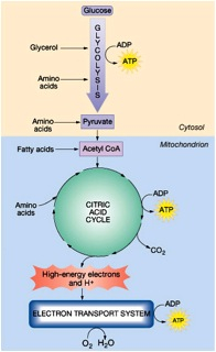
Cats in the wild typically ingest diets containing > 50% of these daily calories as protein (1-3), so that may be a good goal to shoot for when selecting the composition of a diet. When analyzing a diet, I like to examine or calculate the amount of the calories each nutrient (i.e., protein, fat or carbohydrates) provides — this is called the “metabolizable energy,” abbreviated ME (4,5). This measure disregards any part of the food that does not provide any energy (kcal) such as water, ash, or fiber. It only considers the 3 nutrients that provide the needed calories and nothing else.

Let’s take an example using 5 commercial foods containing a wide range of protein content. In each of these diets, I’ve set the fat content and calories (kcal) to be equal, but I’ve varied the protein and carbohydrate amounts. Remember, when we increase or decrease the levels of protein, we must always adjust the levels of carbohydrates, fat, or both to compensate. In other words, the percentage of calories that come protein, fat, and carbohydrates must equal 100% (% protein calories + % fat calories + % carbohydrate calories = 100% of the calories ingested).

So let’s take our first hypothetical diet, which on a dry matter (DM) basis contains 65% protein, 25% fat, and 4% carbohydrate (Table 1). To convert the nutrient composition from a DM to a ME basis, we must remember that protein and carbohydrate both provide approximately 3.5 kcal/g of food, whereas fat provides much more — approximately 8.5 kcal/g of food. Thus, in this diet, the energy provided by protein is 65% times 3.5 kcal/g, or 228 kcal, and the energy provided by fat and carbohydrate are 213 kcal and 14 kcal, respectively, for a total of 454 kcal. The percentage of metabolizable energy that is provided from protein is then calculated (by dividing 228 kcal by 454 kcal and multiplying by 100), which gives us a protein content (ME) of 50%.

Doing the same calculations for diets that contain protein levels (DM basis) of 55%, 45%, 35% and 30% are also included in Table 1. As you can see, looking at nutrients on a dry matter basis “overestimates” the calories provided by protein (ME basis) by ~30%, while greatly underestimating the calories provided by fat (by ~50% in this example).


Table 1. Comparison of Nutrient Content:
Dry Matter (DM) Basis vs. Metabolizable Energy (ME) Basis


Bottom Line: When analyzing the composition of a cat food, one can look at the protein content on a DM or ME basis. In the end, it doesn’t make that much of a difference, but one must realize that the percent protein on a DM basis “overestimates” the calories provided by protein (ME basis). Cats in the wild ingest at least 50% of their calories as protein (ME). As you can see, that equates to ~65% of their diet being composed of protein on a DM basis.

Calculating the Amount of Protein that Commercial Diets Provide

Once we select our diet, we can do the calculations for protein content of a can or cup of food, as I described in detail in my post on “Optimal Protein Requirements for Older Cats and Cats with Hyperthyroidism.” Again, for this calculation, we need to know the protein content of the diet (DM basis), as well as the moisture content of the diet (generally 75% for canned food and 10% for dry food diets).

Let’s take another example by examining 5 commercial foods containing a wide range of protein content (Table 2). All of these diets are 5.5 oz cans (156 g) containing 75% moisture, so this leaves us with 39 g of food in each can on a DM basis (156 times 25% = 39 g). From the information on the label or the company’s website, we will hopefully find the protein content (DM) of each diet (note that if the protein content on a DM basis is not listed, you will have to call the company for that information). The guaranteed analysis (GA) information cannot be used for this calculation, since it listed only the minimum percentage of the crude protein contained the product and is therefore highly inaccurate.

Once the protein content (DM) is know, we multiply that value by total dry matter weight of the can. This provides us with the total protein content in each can of food (e.g., 39 g times 65% = 25.4 g of protein).

In this example, I’ve then used the weight of an average older cat (4.5 kg) to calculate the amount of protein ingested on a body weight basis (g/kg).


Table 2: Calculation of Daily Protein Ingested Based on Protein Content of Food.
*Each 5.5 oz can contains 156 g of food. If moisture content of diet is 75%, that leaves 25% dry matter and 39 g of food (156 times 25% = 39 g).

Bottom Line: As you can see in Tables 1 & 2, we need to feed a diet containing at >50% protein (DM basis) or >40% protein on a ME basis to even come close to providing the needed amounts of protein per kg.

The pet food companies may tell you that these protein levels are unnecessary and that the Association of American Feed Control Officials (AAFCO) requires that feline diets contain a minimum of 26% DM protein for maintenance (4,6). But remember: those are “minimal,” not optimal values. And most importantly, those recommendations are for normal adult cats, not hyperthyroid cats or geriatric cats prone to muscle wasting associated with sarcopenia of aging (7,8).

Is It Even Possible to Feed the Optimal Amounts of Protein to these Geriatic Cats?

The average older cat weighs 10 lbs (4.5 kg); if our goal is to provide 6.0-7.0 g of protein/kg/day, that calculates into an optimal daily protein intake of ~27-31 g. To consume those amounts of protein, that 4.5 cat would have to eat almost three 5.5-oz cans of food per day containing 30% protein (DM), almost two cans of food per day containing 45% protein (DM), or just over one can of a food containing 65% protein (DM) (Table 2).

In general, older cats tend not to eat those large amounts of food each day, unless they have uncontrolled hyperthyroidism and are polyphagic (9,11). Most older euthyroid cats (again weighing 4.5 kg) would eat only one 5.5-oz can per day, which provides only ~2.6 g/kg/day to 5.6 g/kg/day (Table 2).

So what can we do to help prevent "sarcopenia of aging" with its associated muscle wasting in these older cats? We want to feed an energy dense food that contains a highly digestible, high-quality source of protein.

Feeding an Energy-Dense Diet to Older Cats & Cats with Hyperthyroidism

What if our cats won’t eat as much protein as we would like them to ingest?

  1. In addition to providing a diet with an adequate amount of protein, we need to make sure that the cat’s energy requirement is fulfilled. Most of the cat foods marketed as “Senior” diets are too low in caloric content. Remember as cats age (>12 years), their energy requirements actually increase (8, 12-14); these cats will lose weight (and muscle mass) on many of these commercial senior diets.
  2. When calorie intake is inadequate to meet energy needs, body proteins are catabolized and used for energy (4). Again, feeding frequent small meals of an energy-dense, highly digestible diet that meets the senior cat’s increasing energy requirements will minimize protein degradation and avoid protein:calorie malnutrition (8, 12-14).
  3. Identify diets that are palatable for the cat. Many older cats may partially lose their sense of taste or smell. Feeding a variety of different flavors or types of food helps maintain the appetite in many of these older cats.
  4. Warming the food to body temperature or moistening the food may increase help to increase appetite in some cats.
  5. Feeding frequent small meals of energy-dense, fresh food may help increase daily food intake.
  6. Cats are solitary feeders by nature and elderly cats often do not cope well with competition and stressors. Older cats in multi-cat homes may benefit from being fed separately or being offered supplemental meals.
  7. One must also identify underlying medical problems that can lead to decrease in appetite (8,9).

Feeding Highly Digestible, High Quality Protein to Geriatric Cat & Cats with Hyperthyroidism

Again, how can we help compensate for the fact that many older cats will not eat enough food to fulfill their “optimal” daily protein needs? Again, the answer may lie in the source of the protein used in the cat food diet. Feeding a highly digestible, high-quality source of protein would likely allow us to feed lower amounts of protein but still prevent loss of lean body mass and muscle wasting. Improving protein quality without increasing protein intake may help fulfill of these additional protein needs of older cats (4,15).

It is generally accepted that animal protein has a higher digestibility and is of higher quality than that of plant protein sources (16,17). Proteins of plant origin usually have a lower digestibility than animal proteins because plant fiber and the carbohydrates found in plants lower digestion, due to a reduced degradation rate of nutrients in the gut and increased bacterial activity (18).

The biological value (or quality) of a protein is a measure of that protein's ability to supply amino acids (especially the 11 essential amino acids) and to supply these amino acids in the proper proportions (4,15,17). The higher the biological value of a protein, the less would be needed in the diet to meet all of an animal’s essential amino acid requirements.
 

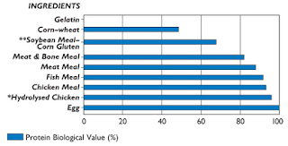
Figure 1: Protein Biologic Value of Common Pet Food Ingredients. 
Data from reference no. 15.

As shown in Figure 1, it is well-established that animal proteins (e.g., meat, meat by-products) have a higher biological values than vegetable proteins (e.g., corn gluten meal, soybean meal, soy protein isolate) (15-17,19). To meet a cat’s optimal daily protein needs, less protein intake would be required when its biological value is high (e.g., when animal protein is fed as the main source of protein).

What About Hill’s y/d? Will Cats Really Eat the Recommended Amounts of y/d Diet Every Day for the Rest of Their Lives ?

Again, one must realize that the amount of protein a cat ingests per day is highly dependent upon the total amount of food consumed. Eating a diet with less-than-optimal quantities of protein might be adequate if that cat ingests a large enough quantity of the food.

The recommended amounts of y/d to feed, as listed on the Hill’s website (20,21), seem reasonable for an uncontrolled hyperthyroid cat. Once euthyroid, however, will the cats continue to consume those same amounts of food? No, that's highly unlikely.  In general, the amount of food hyperthyroid cats ingest decreases as euthyroidism is restored. But remember, older cats still need higher amounts of protein, even when not hyperthyroid, to maintain their muscle mass as they age.

Let’s again take our average older cat weighing 4.5 kg. For that cat to consume protein at our “optimal” amount of 6.0-7.0 g/kg/day, the cat would have to eat almost two 5.5-oz cans of canned Hill's y/d per day or ¾ cup of dry Hill’s y/d per day. Those amounts exceed the amounts listed on the Hill’s feeding guide (18,19). Again, most older euthyroid cats (again weighing 4.5 kg) would eat only one 5.5-oz can per day, which provides ~3.3 g/kg/day, about half of what they actually need.

What about the sources of protein for Hill's y/d diets? Do they include a high-quality, easily digestible source of protein? Are the ingredients of high biological value? Unfortunately, the answer to both questions is no —the ingredients present in y/d are far less than ideal for cats. In addition to the fact that y/d is a low-protein diet, much of the diet’s protein is derived from plant sources. This is especially true for the dry formulation, in which the only listed animal protein on the label is "dried egg product," and this is the fifth ingredient. In other words, this diet does not contain any meat. The primary protein source in dry y/d is corn gluten meal, commonly used in many pet foods because of its low cost.

For the canned formulation, the ingredients list is better in that the first 3 listed ingredients— liver, meat by-products, and chicken—all contain animal protein. Liver is a very nutritious organ meat and a good source of animal protein, but the daily feeding of a pet food containing liver as the first ingredient might be questioned. Do you want to feed your cats a liver diet at every meal for the rest of their lives? Would you consider that a healthy diet?

References

  1. Myrcha A, Pinowski J. Weights, body composition and caloric value of post-juvenile molting European tree sparrows. Condor 1970;72:175–178.
  2. Vondruska JF. The effect of a rat carcass diet on the urinary pH of the cat. Companion Animal Practice 1987;1:5-9.
  3. Crissey SD, Slifka KA, Lintzenich BA. Whole body cholesterol, fat, and fatty acid concentrations of mice (Mus domesticus) used as a food source. Journal of Zoo and Wildlife Medicine 1999;30:222-227. 
  4. Gross KL, Yamka RM, Khoo C, et al. Macronutrients. In: Hand MS, Thatcher CD, Remillard RL, Roudebush R, Novotny, BJ (eds), Small Animal Clinical Nutrition. Mark Morris Institute. 2010; 49-105. 
  5. Schenck PA. Nutrients. In: Home-Prepared Dog and Cat Diets.  Wiley-Blackwell. Ames IA, 2010; 23-49.
  6. AAFCO. (Association of American Feed Control Officials). Official Publication, 2007. 
  7. Wolfe RR. Sarcopenia of aging: Implications of the age-related loss of lean body mass. Proceedings of the Nestlé Purina Companion Animal Nutrition Summit: Focus on Gerontology. St. Louis, MO. 2010, pp. 12-17. 
  8. Little S: Evaluation of the senior cat with weight loss, In: Little, S. (ed), The Cat: Clinical Medicine and Management. Philadelphia, Elsevier Saunders, in press. 
  9. The Special Needs of the Senior Cat. Information brochure, Cornell University College of Veterinary Medicine.   
  10. Peterson ME, Kintzer PP, Cavanagh PG, et al. Feline hyperthyroidism: pretreatment clinical and laboratory evaluation of 131 cases. Journal of the American Veterinary Medical Association 1981;183:103-110. 
  11. Broussard JD, Peterson ME, Fox PR. Changes in clinical and laboratory findings in cats with hyperthyroidism from 1983 to 1993. Journal of the American Veterinary Medical Association 1995;206:302-305. 
  12. Perez-Camargo G: Cat nutrition: What is new in the old? Compendium for Continuing Education for the Practicing Veterinarian 2004;26 (Suppl 2A):5-10.
  13. Pérez-Camargo G. Feline decline in key physiological reserves: implication for mortality. Proceedings of the Nestlé Purina Companion Animal Nutrition Summit: Focus on Gerontology. St. Louis, MO. 2010, pp. 6-13. 
  14. Sparkes AH. Feeding old cats— An update on new nutritional therapies. Topics in Companion Animal Medicine 2011;26:37-42. 
  15. Lewis LD, Morris ML, Hand MS. Nutrients. In: Small Animal Clinical Nutrition III. Mark Morris Associates. Topeka, 1987; 1-25.
  16. Funaba M, Matsumoto C, Matsuki K, et al. Comparison of corngluten meal and meat meal as a protein source in dry foods formulated for cats. American Journal of Veterinary Research 2002;63:1247-1251.
  17. Funaba M, Oka Y, Kobayashi S, et al. Evaluation of meat meal,chicken meal, and corn gluten meal as dietary sources of protein in dry catfood. Canadian Journal of Veterinary Research 2005;69:299-304.
  18. Murray SM, Patil AR, Fahey GC, et al. Raw and rendered animal by-products as ingredients in dog diets. Journal of Animal Science 1997; 75:2497-2505. 
  19. Feline Nutrition. http://maxshouse.com/feline_nutrition.htm
  20. Hill's Pet Nutrition website. Prescription Diet y/d Thyroid Feline Health (Dry).
  21. Hill's Pet Nutrition website. Prescription Diet y/d Thyroid Feline Health (Canned).

Źródło: endocrinevet.blogspot.com

As I discussed in a recent post on Optimal Protein Requirements for Older Cats and Cats with Hyperthyroidism, energy requirements sharply and progressively increase in older cats starting at 10 to 12 years of age (1-3). If daily caloric intake is not increased, progressive weight loss will result, due in large part to the loss of lean body mass (i.e., muscle mass), a phenomenon referred to “sarcopenia” of aging (4-7).

In addition to this increased caloric intake, older cats also require higher amounts of protein to maintain protein reserves compared with younger adult cats (3, 8-11). As cats age, they absorb and metabolize protein less efficiently (10). Therefore, it’s extremely important to feed high-quality protein (i.e., animal source rather than grain-based), as well as an adequate quantity of protein to aging cats.

Animals derive energy from the oxidation of the macronutrients carbohydrate, fat, and protein (12). Most animals, including rats and humans, generally adapt to the diet being fed and can oxidize whatever fuel mixture is contained in their prevailing diet (13,14). This enables most animals (especially omnivores) to use widely differing diet compositions to satisfy their energy requirements.

A number of veterinarians have contacted me over the last few weeks with concerns about the potential problems associated with feeding a high protein diet to the geriatric cat, especially in those cats with kidney disease. Basically, the questions come down to the following:

  • If animals can adapt their energy intake based on a wide variety of feeding regimens, why can’t we just increase the amounts of fat and carbohydrate fed to compensate for the increasing energy requirements in our senior (and hyperthyroid) cats? Won't that help prevent loss of muscle mass?
  • But can cats as obligate carnivores adapt to the same extent?

In this post, my mission is to review energy and protein metabolism in cats. I will attempt to explain why, unfortunately, increasing the amount of fat or carbohydrate fed to an older or hyperthyroid cat generally cannot compensate for a diet deficient in an optimal protein content.

Overview of the Body's Energy Production (Nutritional Biochemistry 101)


Fig. 1: Pathways of
intermediary metabolism

Let's start with a brief review of nutritional biochemistry. If you are a practicing veterinarian like myself, it's highly likely that you may not remember all of the metabolic pathways an animal uses for energy production.

The initial biochemical reactions by which energy is derived from carbohydrates, fats, and proteins are different. However, all 3 macronutrients eventually go through a final common pathway for energy generation called the citric acid cycle — also known as the tricarboxylic acid cycle (TCA) cycle or the Krebs cycle (Figure 1) (12,15).

Carbohydrates: Glucose derived from dietary carbohydrates is first oxidized through the glycolysis pathways to yield pyruvate and then acetyl-CoA. The acetyl-CoA is then oxidized in the citric acid cycle (12,15). High-energy electrons produced in the citric acid cycle enter the electron transport chain to generate adenosine triphosphate (ATP), which transfers its energy from chemical bonds to energy-absorbing chemical reactions within the cell (Figure 1).

Fats: Fatty acids from dietary fats are initially oxidized to acetyl-CoA by the beta-oxidation pathway, which then enter the citric acid cycle and subsequently generate ATP via oxidative phosphorylation in the electron transport system (Figure 1).


Figure 2: Protein catabolism

Protein: Once protein synthesis is maximized, excess amino acids can be used for energy by undergoing transamination and deamination. In oxidative deamination, an amino group is removed from the amino acid and converted to ammonia.

The remaining carbon skeleton from these deaminated amino acids (organic ketoacids) can be recycled to make nonessential amino acids, or they can be oxidized for energy (12,15). If used for energy, the catabolized amino acids enter either the glycolysis pathway or the citric acid cycle to ultimately form ATP in the electron transport system (Figure 1). The carbon skeletons of catabolized amino acids can also be converted to glucose or ketone bodies in the liver.

Ammonia is toxic to the body, so enzymes convert it to urea by addition of carbon dioxide in the urea cycle, which takes place in the liver. Urea can safely diffuse into the blood and then be excreted into the urine (Figure 2).

Interconversion of nutrient molecules: In metabolism, there commonly is interconversion of nutrient molecules. Excess glucose is stored as glycogen (glycogenesis); when glycogen stores are filled, glucose and amino acids are used to synthesize lipids (lipogenesis). Therefore, glycogen and adipose tissue both serve as long-term forms of energy storage in the body.

In contrast, amino acids are not "stored" in the body, other than that found in muscle and other structural proteins. Although amino acids can be converted to either fat or glucose, the opposite does not occur — fat and carbohydrates cannot be directly converted to amino acids to be made into protein (12-15).

Protein Structure, Functions & Metabolism

Proteins, from the Greek proteios meaning "first" (16), are important biological molecules that consist of string of amino acids linked together in sequence as polypeptide chains. Proteins vary in shape and size, some consisting of only 20-30 amino acids and others of several thousands (12,15).

Proteins are present in every living cell. In the skin, hair, cartilage, muscles, tendons, and ligaments, proteins hold together, protect, and provide structure to the body. As enzymes, hormones, antibodies, and globulins, they catalyze and regulate body chemistry. In addition, protein is required for growth and tissue repair, as well as for maintaining muscle mass and tone.

Essential and Nonessential Amino Acids
More than 20 amino acids are involved in the synthesis of protein in the body. Essential amino acids are those that cannot be formed in sufficient amounts to meet the requirements for growth and maintenance and must be supplied in the diet. Nonessential amino acids are those that the body can produce in sufficient amounts from other nutrients and metabolites and, thus, do not need to be supplied in the diet.

Even with the nonessential amino acids, however, fat and carbohydrates cannot be directly converted to amino acids to be made into protein (12,15). Although all mammals can synthesize 10 nonessential amino acids from precursor carbon skeletons (if adequate nitrogen and energy are available), there is no direct way that amino acids can be synthesized directly from either fat or carbohydrates.

Amino Acid Catabolism & Synthesis—Protein Turnover
Although essential amino acids are not stored as such in the body for any significant period of time, all body proteins are continuously being broken down and resynthesized in a process known as protein turnover (12,15,17). During protein turnover, some amino acids enter catabolic pathways and are permanently lost. Therefore, adequate amounts of high-quality dietary protein (containing all the essential amino acids) must be consumed each day to replace the both the essential and nonessential amino acids lost to catabolism.

Hypermetabolic states, such as hyperthyroidism or other illness, increase both protein turnover and nitrogen losses and therefore increase the daily protein requirements (18,19).

Protein as an Energy Source
Unlike fat or carbohydrate, protein cannot be stored as such in the body (other than as muscle protein itself).  In animals fed diets containing more protein than is needed, the extra protein is metabolized and used for energy (Figures 1 & 2).

In all species, but most importantly in cats (see below), ingested amino acids are converted to carbohydrates via gluconeogenesis (12,15,17, 20-22). This pathway is also used under starvation conditions to generate glucose and energy from the body's own proteins, particularly those found in muscle.

Factors That Determine How Dietary Proteins are Used

  • All or None Rule: To make a certain protein, the necessary amino acids must be present in the cell in the right amounts, or the protein will not be made. Essential amino acids that are not used to make proteins are not stored.
  • Adequacy of Caloric Intake:  If the diet fails to provide sufficient calories as carbohydrates and fats, proteins will not be synthesized; instead the ingested amino acids will be used as a source of energy.
  • Nitrogen Balance:  Normally, the amount of protein synthesis is equal to the amount of protein breakdown (12,17). If there is more protein synthesis than breakdown, then we have a positive nitrogen balance (e.g., recovery from injury). If there is greater protein breakdown than synthesis, then there is a negative nitrogen balance (e.g, starvation, illness, hyperthyroidism).
  • Hormonal Controls:  Anabolic hormones (e.g., insulin, growth hormone, sex hormones) stimulate the production or maintenance of proteins.  Catabolic hormones (e.g, glucocorticoids, thyroid hormone) stimulate the breakdown of proteins (18,19).

Cats Do Not Need Carbohydrate But Have High Protein Requirements

The cat, as a strict carnivore, has evolved to to depend on protein as a major energy source. The "natural" diet of cats in the wild is based upon the consumption of small mammals, birds, and insects — the composition of this diet is high in protein and fat but low in carbohydrate (20-22). This natural diet high in animal protein contains all of a cat's essential amino acids, which is not the case for plant-based proteins.

Cats have no dietary requirement or need for carbohydrate but are adapted physiologically and metabolically for high protein intake (23). In support of that, normal cats do well when fed diets containing 70% protein (24-27). The lack of a need for dietary carbohydrates is related to the fact that cats have developed a tremendous ability to synthesize their needed glucose from protein catabolism via hepatic gluconeogenesis (24,25,28).

Cats have a much higher protein requirement than other species, such as dogs and humans. The protein requirement for the adult cat is 2-3 times as much as the adult dog (23), and their protein requirement increases even further as cats reach old age (3,8-11). This high protein requirement of cats is primary related to the fact that cats have a limited ability to decrease the hepatic enzymes responsible for amino acid catabolism, even when fed a lower than optimal protein intake (24,29,30).

The Domestic Cat: A Metabolically Inflexible Carnivore

When most omnivores (e.g., humans, rats, pigs, dogs), ingest a diet high in protein, the activities of the amino acid-catabolizing enzymes in the liver increase to cope with the higher flux of amino acids (31-33). The activity of the urea cycle enzymes also increase to metabolize the increased ammonia generated upon the catabolism of the amino acids. On the other hand, when fed a diet low in protein, omnivores accommodate by lowering the hepatic activity of these catabolic enzymes of amino acid metabolism, as well as decreasing hepatic urea production (31-33).

By contrast, cats have a very limited ability to down-regulate these hepatic catabolic enzymes when fed a low-protein diet. In a classic study, Rogers et al (29) compared the activity of several catabolic enzymes of amino acid metabolism in adult cats fed either a high- or low-protein diet or fasted for 5 days. Results showed little changes in the hepatic enzyme activities between the 3 groups of cats, with hepatic enzyme activities remaining set at high levels to cope with a high protein diet (even when they weren’t being fed!).

Why would cats be so different?
Well, compared to other carnivores, cats may not be so strange after all. A similar degree of metabolic "inflexibility" has also been reported in other carnivores such as barn owls and the rainbow trout (34,35).

This limited metabolic flexibility in cats and other obligate carnivores likely represents an evolutionary adaptation to a consistently abundant supply of dietary protein. The moderately high and fixed activity of the urea cycle provides a safeguard against ammonia toxicity after ingestion of a high-protein meal. In addition, the high rate of amino acid catabolism allows for a readily available source of energy via direct oxidation or as a substrate for hepatic gluconeogenesis.

It is only when a cat is fed a lower protein diet, a condition that would never happen in the wild, that the high rate of protein catabolism becomes a disadvantage.

Bottom Line

  • Unlike omnivores (dogs, pigs, rats, humans), cats have a limited ability to decrease the activity of the hepatic enzymes responsible for removing amino groups from the amino acids when fed a low protein diet. 
  • Because these feline hepatic enzyme systems are constantly active, a fixed amount of dietary (or muscle) protein will always be catabolized for energy no matter how much energy in the form of carbohydrate or fat the cat ingests. 
  • In addition, neither fat nor carbohydrates can be directly converted to amino acids to be made into protein. In this regard, the carnivorous cat is similar to omnivores.
  • Overall, this explains why muscle wasting can occur so quickly in the older, geriatric cat, which becomes ill or develops a poor appetite or is fed a low-protein diet.

References:

  1. Perez-Camargo G: Cat nutrition: What is new in the old? Compendium for Continuing Education for the Practicing Veterinarian 2004;26 (Suppl 2A):5-10.
  2. Laflamme D. Nutrition for aging cats and dogs and the importance of body condition. Veterinary Clinics of North America: Small Animal Practice 2005;35:713-742.
  3. Pérez-Camargo G. Feline decline in key physiological reserves: implication for mortality. Proceedings of the Nestlé Purina Companion Animal Nutrition Summit: Focus on Gerontology. St. Louis, MO. 2010, pp. 6-13.
  4. Fujita S, Volpi E. Nutrition and sarcopenia of ageing. Nutrition Research Reviews 2004;17:69-76.
  5. Paddon-Jones D, Short KR, Campbell WW, et al. Role of dietary protein in the sarcopenia of aging. The American Journal of Clinical Nutrition 2008;87:1562S-1566S. 
  6. Short KR, Nair KS. Mechanisms of sarcopenia of aging. Journal of Endocrinological Investigation 1999;22(5 Suppl):95-105.
  7. Wolfe RR. Sarcopenia of aging: Implications of the age-related loss of lean body mass. Proceedings of the Nestlé Purina Companion Animal Nutrition Summit: Focus on Gerontology. St. Louis, MO. 2010, pp. 12-17.
  8. Little SE: Evaluation of the senior cat with weight loss, In: Little, S.E. (ed), The Cat: Clinical Medicine and Management. Philadelphia, Elsevier Saunders, 2012;1176-1181.
  9. Sparkes AH. Feeding old cats— An update on new nutritional therapies. Topics in Companion Animal Medicine 2011;26:37-42.
  10. Patil AR, Cupp C, Pérez-Camargo G. Incidence of impaired nutrient digestibility in aging cats. Nestlé Purina Nutrition Forum Proceedings. 2003;26,2(A):72.
  11. Wakshlag JJ. Dietary protein consumption in the healthy aging companion animal. Proceedings of the Nestlé Purina Companion Animal Nutrition Summit: Focus on Gerontology. St. Louis, MO. 2010, pp. 32-39.
  12. Gross KL, Yamka RM, Khoo C, et al. Macronutrients. In: Hand MS, Thatcher CD, Remillard RL, Roudebush R, Novotny, BJ (eds), Small Animal Clinical Nutrition. Mark Morris Institute. 2010; 49-105. 
  13. Davy KP, Horton T, Davy BM, et al. Regulation of macronutrient balance in healthy young and older men. International Journal of Obesity and Related Metabolic Disorders 2001;25:1497-1502.
  14. Galgani J, Ravussin E. Energy metabolism, fuel selection and body weight regulation. International Journal of Obesity 2008;32 (Suppl 7):S109-119.  
  15. Brody T. Nutritional Biochemistry. Second Edition. Academic Press. 1998.
  16. New Oxford Dictionary of English. Oxford University Press, 2001.
  17. Berg JM, Tymoczko JL, Stryer L. Protein turnover and amino acid catabolism. Biochemistry. 5th edition ed. New York: W H Freeman, 2002. 
  18. Mitch WE. Mechanisms accelerating muscle atrophy in catabolic diseases. Transactions of the American Clinical and Climatological Association 2000;111:258-269. 
  19. Riis AL, Jørgensen JO, Gjedde S, et al. Whole body and forearm substrate metabolism in hyperthyroidism: evidence of increased basal muscle protein breakdown. American Journal of Physiology: Endocrinology and Metabolism 2005; 288:E1067-1073. 
  20. Myrcha A, Pinowski J. Weights, body composition and caloric value of post-juvenile molting European tree sparrows. Condor 1970;72:175–178.
  21. Vondruska JF. The effect of a rat carcass diet on the urinary pH of the cat. Companion Animal Practice 1987;1:5-9.
  22. Crissey SD, Slifka KA, Lintzenich BA. Whole body cholesterol, fat, and fatty acid concentrations of mice (Mus domesticus) used as a food source. Journal of Zoo and Wildlife Medicine 1999;30:222-227. 
  23. National Research Council. Proteins and amino acids. In: Nutrient Requirements of Dogs and Cats. Washington, DC: National Academies Press. 2006; 111-145.
  24. MacDonald ML, Rogers QR, Morris JG. Nutrition of the domestic cat, a mammalian carnivore. Annual Review of Nutrition 1984;4:521-562. 
  25. Morris JG. Idiosyncratic nutrient requirements of cats appear to be diet-induced evolutionary adaptations. Nutrition Research Reviews 2002;15:153-168. 
  26. Hendriks WH. Protein metabolism in the adult domestic cat (Felis catus). PhD Thesis, 1996. 
  27. Hamper B, Bartges J, Kirk C, et al. The unique nutritional requirements of the cat: a strict carnivore In: Little SE, ed. The Cat: Clinical Medicine and Management. St. Louis: Elsevier Saunders, 2012;236-242.
  28. Kettelhut IC, Foss MC, Migliorini RH. Glucose homeostasis in a carnivorous animal (cat) and in rats fed a high-protein diet. American Journal of Physiology 1980; 239:R437-440.
  29. Rogers QR, Morris JG, Freedland RA. Lack of hepatic enzymatic adaptation to low and high levels of dietary protein in the adult cat. Enzyme 1977;22:348-356.
  30. Green AS, Ramsey JJ, Villaverde C, et al. Cats are able to adapt protein oxidation to protein intake provided their requirement for dietary protein is met. The Journal of Nutrition 2008;1053-1060. 
  31. Waterlow JC. The mysteries of nitrogen balance. Nutrition Research Reviews 1999;12:25-54. 
  32. Rosebrough RW, Steele NC, McMurtry JP. Effect of protein level and supplemental lysine on growth and urea cycle enzyme activity in the pig. Growth 1983;47:348-260.
  33. Schimke RT. Adaptive characteristics of urea cycle enzymes in the rat. Journal of Biological Chemistry  1962;237:459-68. 
  34. Walton MJ. Metabolic effects of feeding a high protein/low carbohydrate diet as compared to a low protein/high carbohydrate diet to rainbow trout Salmo gairdneri. Fish Physiology and Biochemisty 1986;1;7-15. 
  35. Meyers MR, Klasing KC. Low glucokinase activity and high rates of gluconeogenesis contribute to hyperglycemia in barn owls (Tyto alba) after a glucose challenge. Journal of Nutrition 1999; 129:1896-1904.

Źródło: endocrinevet.blogspot.com

My problem case is an 14-year old, male-neutered DLH cat that has been on a relatively high dose of lactulose (6 ml, bid) for chronic constipation for many years.

One month ago, he developed weight loss, an increase in appetite, and polydipsia. We diagnosed diabetes mellitus (he had severe hyperglycemia with glucosuria) and started him on insulin glargine (Lantus, Sanofi-Aventis). The owner has kept the cat on Hill's dry Science Diet, which the cat loves.

So far, the cat's glargine insulin dose is up to 4 U twice a day (0.9 units/kg), but he is not yet showing any improvement. His last blood glucose was still high at 381mg/dl, with marked glucosuria but negative ketonuria. A serum fructosamine was high at 630 μmol/L (normal <450 μmol/L). A blood glucose curve demonstrated persistent hyperglycemia throughout the day.

My questions are are 4-fold:

  1. Does this cat have insulin resistance?
  2. Would lactulose have any bearing on this high of blood glucose? Should I try stopping it to see if hyperglycemia improves on insulin therapy?
  3. Should I change his insulin to PZI (ProZinc; Boehringer Ingelheim) or another insulin analogue?
  4. Is his present diet acceptable?

My Response:

Lactulose is a synthetic, nondigestible sugar developed as a treatment for chronic constipation. It is a disaccharide formed from one molecule each of the monosaccharides fructose and galactose (see Figure 1).

Because it is not digested, lactulose passes unchanged to reach the colon, where it exerts its osmotic laxative effect.Therefore, the lactulose did not contribute to your cats diabetes since the "sugar" in lactulose is not absorbed. Similarly, the drug would not be an issue contributing to the lack of response to insulin therapy in this cat.


Structure of lactulose, a disaccharide

Recommended Steps in this Cat's Management:

1. Rule out urinary tract infection and other concurrent problems

  • To start, I'd recommend a urine culture to rule out an occult urinary tract infection. Cat with diabetes are prone to developing urinary tract infections (UTI), so periodic urine cultures are a good idea.

2. Change to a high-protein, low-carbohydrate diet

  • You should also consider switching this cat's diet to a good canned food diet — one with a composition that is low in carbohydrates (<10% of calories) and higher in protein (>40% of calories). By lessening the insulin resistance that is a hallmark of diabetes in cats, that diet change will make cats more sensitive to the effects of insulin. 
  • Most of my diabetic cats are doing well on an over-the-counter canned food that are low enough in carbohydrates and high in protein content. Check out this website (binkyspage.tripod.com/canfood), which gives you a breakdown of the composition of the various prescription and over-the-counter diets. It turns out that many of the over-the-counter diets have a better composition of protein and carbohydrates than you might have thought — even better than many of the more expensive prescription diets. Very few of my diabetic cat patients require a prescription diet to fulfill their nutritional needs.
  • Almost all dry food cat diets are much too high in carbohydrates and  too low in protein content. That is why I believe it's best to limit the amount of dry food that is fed to diabetic cats, or even better, not feed dry food at all.

3. Consider a change in insulin preparation

  • Once you have excluded UTI as a cause of insulin resistance, I would change to a more appropriate "diabetic" diet. If no improvement in insulin dosage or glucose regulation is noted after 2-4 weeks, I would recommend switching from insulin glargine to another insulin preparation.
  • If needed, I'd recommend a change to PZI (ProZinc), starting at a lower initial dose (e.g., 2 U, BID), and doing dose regulation as needed. I've now seen a few cats that could not be regulated on glargine do better on this insulin preparation.

References:

  1. Rucinsky R, Cook A, Haley S, Nelson R, et al. AAHA diabetes management guidelines for dogs and cats. Journal of the American Animal Hospital Association 2010;46:215-224.
  2. Frank G, Anderson W, Pazak H,  et al. Use of a high-protein diet in the management of feline diabetes mellitus. Veterinary Therapeutics 2001;2:238-246.
  3. Rand JS, Fleeman LM, Farrow HA, eet al. Canine and feline diabetes mellitus: nature or nurture? The Journal of 2004;134(8 Suppl):2072S-2080S.
  4. Nelson RW, Henley K, Cole C, et al. Field safety and efficacy of protamine zinc recombinant human insulin for treatment of diabetes mellitus in cats. Journal of Veterinary Internal Medicine 2009;23:787-793.

Źródło: endocrinevet.blogspot.com

What form would Liothyronine sodium be given for a T3 suppression test?

What testing protocol do you use? Can you explain how you interprete the test results?

My Response:

Liothyronine sodium is the L-isomer of triiodothyronine (T3). I have always used tablets that contain 25 μg. The common trade name for L-T3 is Cytomel (Figure 1).

Protocol for T3 suppression test in cats

The test protocol that I developed over 20 years ago and still use is as follows (1):

Figure 1: Cytomel
  1. One day 1, draw a blood sample is drawn for determination of baseline serum concentrations of total T4 and T3. This serum sample is not yet submitted to the laboratory but kept refrigerated (or frozen) until day 4.
  2. Owners are instructed to give 7 doses of a T3 pill (liothyronine sodium; Cytomel) to their cat, beginning on the following morning.
  3. On day 2 and 3, the owners administer the liothyronine at a dosage of 25 µg every 8 hours for 2 days (6 doses).
  4. On the morning of day 4, a seventh 25-µg dose of liothyronine is given and the cat returned to the veterinary clinic within 2 to 4 hours.
  5. The veterinarian again draws a blood sample for serum T4 and T3 determinations.
  6. Both the basal (day 1) and post-liothyronine (day 4) serum samples are submitted to the laboratory together to eliminate the effect of between assay variation in hormone concentrations.
Figure 2: T3 suppression tests in normal cats (left) & cats with hyperthyroidism (right)

Interpretation of results of T3 suppression test

Regarding interpretation of T3 suppression test results, I find that the absolute serum T4 concentration after liothyronine administration is the best means of distinguishing hyperthyroid cats from normal cats or cats with nonthyroidal disease (1-5).

Cats with hyperthyroidism have post-liothyronine serum T4 values greater than 20 nmol/L (greater than 1.5 μg/dl), whereas normal cats and cats with nonthyroidal disease have T4 values less than 20 nmol/L (Figure 2). There may be a great deal of overlap of the per cent decrease in serum T4 concentrations after liothyronine administration between the three groups of cats, but suppression of 50 per cent or more only occurs in cats without hyperthyroidism.

Serum T3 concentrations, as part of the T3 suppression test, are not useful in the diagnosis of hyperthyroidism per se. However, these basal and post-liothyronine serum T3 determinations can be used to monitor owner compliance with giving the drug. If inadequate T4 suppression is found, but serum T3 values do not increase after treatment with liothyronine, problems with owner compliance should be suspected and the test result considered questionable.

Disadvantages of the T3 suppression test

Overall, the T3 suppression test is very useful for diagnosis of mild hyperthyroidism in cats, but the test does come with disadvantages:

  • T3 suppression testing is a relatively long test (3 days)
  • Owners are required to give multiple doses of liothyronine
  • Cats must swallow the tablets if the test is going to be meaningful.
  • If the liothyronine is not administered properly or the cat does not swallow the liothyronine tablet, circulating T3 concentrations will not rise to decrease pituitary TSH secretion (Figure 2), and the serum T4 value will not be suppressed, even if the pituitary-thyroid axis is normal. Failure of a cat to ingest the liothyronine could result in a false-positive diagnosis of hyperthyroidism in a normal cat or cat with nonthyroidal disease.

References:

  1. Peterson ME, Graves TK, Gamble DA: Triiodothyronine (T3) suppression test. An aid in the diagnosis of mild hyperthyroidism in cats. Journal of Veterinary Internal Medicine 1990;4:233-238.
  2. Peterson ME. Diagnostic tests for hyperthyroidism in cats. Clinical Techniques in Small Animal Practice 2006;21:2-9.
  3. Peterson ME: Diagnostic testing for feline hyper- and hypothyroidism. Proceedings of the 2011 American College of Veterinary Internal Medicine (ACVIM) Forum. pp. 95-97, 2011
  4. Peterson ME: Hyperthyroidism in cats, In: Rand, J (ed), Clinical Endocrinology of Companion Animals. New York, Wiley-Blackwell, in press.
  5. Mooney CT, Peterson ME: Feline hyperthyroidism, In: Mooney CT, Peterson ME (eds), Manual of Canine and Feline Endocrinology (Fourth Ed), Quedgeley, Gloucester, British Small Animal Veterinary Association, 2012; in press.

Źródło: endocrinevet.blogspot.com

Scientific Paper Review— Facing the Nuclear Threat: Thyroid Blocking Revisited

Hänscheid H, Reiners C, Goulko G, Luster M, Schneider-Ludorff M, Buck AK, and Lassmann M. Facing the nuclear threat: thyroid blocking revisited. Journal of Clinical Endocrinology and Metabolism 2011;96:3511-3516.
 

Two common preparations of Potassium Iodide available in the US

Summary of Paper

Background:
Radioiodine-131 is a major fission product released into the atmosphere after a nuclear accident and results in contamination of water and soil. It may be ingested through food and water that is contaminated with 131-iodide. The most recent accident, of course, was the nuclear reactor damage from the Tsunami in Japan.

Radioidine-131 from the Chernobyl accident resulted in a dramatic increase of  thyroid cancer in children who were exposed to the radiation. To prevent thyroid  cancer from thyroid uptake of 131-iodide, the World Health Organization and the U.S. National Research Council have recommended that the potentially exposed population be given tablets containing 100 mg of iodide as potassium iodide to block the uptake of the radioiodide (1).

Potassium iodide is the same form of iodine used to iodize table salt. Potassium iodide floods the thyroid with iodine, thus preventing radioactive iodine from being absorbed (2). Taking potassium iodide immediately after a nuclear accident appears to lessen the risk of developing thyroid cancer.

Sodium perchlorate is a chemical that causes the thyroid to release any iodine that is stored in the thyroid cells (3). Thus, taking sodium perchlorate after a nuclear accident may cause the thyroid to release any radioactive iodine that the thyroid cells have already taken up.

This study was done to see if taking sodium perchlorate might also lower the chance of thyroid cancer after a nuclear accident. To that end, the effectiveness of sodium perchlorate as a blocking agent was compared with the use of potassium iodide in patients exposed to radioactive iodine.

Methods:
Twenty-seven healthy euthyroid subjects with a mean age of 25 years participated in 48 studies of 123-I kinetics in the thyroid. None of these patients were known to have any thyroid disease.

Each volunteer received a small tracer dose of radioactive iodine (I-123), then different amounts of either potassium iodide or sodium perchlorate. The volunteers then had a radioactive iodine uptake (RAIU) test to see how long the radiation stayed in the thyroid.

Results:
The authors found that 100 mg of either potassium iodide or sodium perchlorate was able to reduce the thyroid absorbed dose of radioactive iodine by almost 90%.

Iodine kinetics were significantly faster in subjects younger than 25 years of age, as compared with those older than 25 years. The time of intervention to achieve a 50% dose reduction was 2.4 hours fort he person with the fastest kinetics and 9.2 hours for the subject with the slowest iodine kinetics.

Conclusions: 
At a time of a nuclear disaster, shielding from radiation by distance, staying indoors, and taking all other measures to avoid exposure to radiation are important. Public authorities must act quickly to distribute iodide tablets to the population who may be exposed in order to prevent thyroid uptake of 131-I that has strong beta radiation, which can induce DNA damage and result in thyroid cancer (1). The data of this paper show that the best blockade occurs when the blocking agent is given before the patient is exposed to radioiodine.

Currently, potassium iodide is the main compound to take after a nuclear accident and is stockpiled in areas that have nuclear power plants in the United States. The results suggest that both potassium iodide and sodium perchlorate lower radioactive iodine levels to the thyroid after a nuclear accident. Both agents are relatively safe to take as a single dose following a nuclear accident.

Since thyroid effects of low levels of perchlorate in the U.S. environment are controversial, however, it is unlikely that it will be made available to the general U.S. public following a nuclear accident. However, it is another compound that can be used in this situation.

Current guidelines for blocking 131- I uptake are adequate for older individuals but probably overestimate the efficacy of blocking in young people, who have faster kinetics. Perchlorate may be used for thyroid blocking as an alternative for individuals with iodine hypersensitivity or those at risk for iodine-induced hyperthyroidism. The conclusion that thyroid iodine kinetics vary among individuals is not novel, but the point that younger people have faster turnover and may need a larger blocking dose given at an earlier time is important.

Implications of the Study (including Protecting our Dogs and Cats)

What about our companion animals? The iodine kinetics for both dogs and cats tends to be more rapid than in human patients, which could suggest that larger doses of potassium iodide or perchlorate might be needed in case of an emergency.

In one study of normal dogs, the blocking action of both potassium iodide and potassium perchlorate was about 90%, similar to the findings of this present study (5). Potassium iodide was chosen for its limited side effects and more universal utilization. In that canine study, results indicated that 25 mg of potassium iodide is the ideal amount to be administered to the dog. This corresponds to approximately 100 mg for a 70 kg human being (i.e., a dose of 1.4 mg/kg).

For more information about use of potassium iodide in dogs and cats, see my related post on "Radiation Toxicity, Potassium Iodide, and Our Pets."

References:

  1. National Research Council. Distribution and  administration of potassium iodide in the event of a nuclear incident, Washington, DC: National Academies Press, 2004.
  2. Sternthal E, Lipworth L, Stanley B, Abreau C, Fang SL, Braverman LE. Suppression of thyroid radioiodine uptake by various doses of stable iodide. New England Journal of Medicine 1980;303:1083-1088.
  3. Greer MA, Goodman G, Pleus RC, Greer SE. Health effects assessment for environmental perchlorate contamination: the dose response for inhibition of thyroidal radioiodine uptake in humans. Environmental Health Perspectives 2002;110:927-937.
  4. Ribela MT, Marone MM, Bartolini P. Use of radioiodine urinalysis for effective thyroid blocking in the first few hours post exposure. Health Physics 1999;76:11-16.

Źródło: endocrinevet.blogspot.com

3

bony.jpg

zoetis2024

megavet