")

Addison's disease, the common name for hypoadrenocorticism or adrenal insufficiency, as I have discussed in my last two blog posts, is a disease with vague clinical features that are common in many other ailments, making diagnosis difficult in many cases. But once Addison's disease is correctly diagnosed, a properly treated dog can live a normal and happy active life.
 The missing adrenal hormones
The adrenal, one on each kidney, is made up of two layers, the cortex and the medulla. The inner medulla secretes epinephrine (adrenaline) and is not affected by Addison's disease. The outer cortex layer secretes two corticosteroid hormones, cortisol and aldosterone, both of which are deficient in Addison's disease.
Aldosterone is a corticosteroid hormone (more specifically, a mineralocorticoid — think minerals: salt, sodium, potassium) responsible for maintaining normal circulating electrolyte levels. Once secreted, aldosterone acts on the kidney to conserve sodium, excrete potassium, and retain needed water.
Cortisol is also a corticosteroid hormone (in this case, a glucocorticoid — think glucose, sugar, energy) that is essential for life. It supports a variety of important cardiovascular, metabolic, immunologic, and stress functions.
Not all forms of hypoadrenocorticism are treated the same
There are three forms of hypoadrenocorticism: primary, secondary and atypical Addison's disease.
- Primary Addison's disease is most commonly is the result of immune-mediated damage to the glands.
- Atypical Addison's disease is a poorly understood disorder, thought to generally be an early stage of primary Addison's disease.
- Secondary hypoadrenocorticism results from a deficiency of the pituitary hormone, adrenocorticotropic hormone (ACTH). Without circulating ACTH, cortisol cannot be secreted by the adrenal glands.
It is important which form of hypoadrenocorticism is present in order to provide the correct treatment. In primary hypoadrenocorticism, both cortisol and aldosterone are deficiency and must be replaced for life. In atypical and secondary hypoadrenocorticism, on the other hand, only the glucocorticoids need to be replaced, at least initially.
Treating the acute adrenal crisis: A true medical emergency
Untreated, hypoadrenocorticism (especially primary Addison's disease) can lead to an adrenal crisis. An adrenal crisis is a medical emergency that requires intravenous fluids and glucocorticoids to restore the body’s levels of fluids, salt, and sugar to normal.
Once stabilized, the dog can then be treated with glucocorticoid and mineralocorticoid replacement therapy at home.
Treating chronic hypoadrenocorticism: A lifelong disease
Depending of the subtype of hypoadrenocorticism, synthetic corticosteroid drugs that act like mineralocorticoid or glucocorticoids used for hormone replacement therapy.
Mineralocorticoid treatment
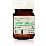
For mineralocorticoid (aldosterone) replacement, either an oral medication called fludrocortisone acetate (Florinef™) or the injectable desoxycorticosterone pivalate (DOCP; Percorten-V™) is used.
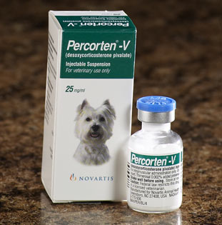 We typically institute treatment with DOCP (Percorten-V) at a dosage of 2.2 mg/kg, subcutaneously or intramuscularly, every 25 to 30 days. Side effects associated with DOCP therapy are rare. This dosage interval is effective in almost all dogs, and most are well controlled with a DOCP injection every 4 weeks.
Initially, serum kidney and electrolyte concentrations should be monitored at approximately 2-weeks intervals in order to determine the drug’s peak effect and to help make necessary dosage adjustments. Once stabilized, serum electrolyte and creatinine concentrations are checked every 3 to 6 months. Because DOCP is a pure mineralocorticoid and has no glucocorticoid activity, it is essential that dogs receive concurrent glucocorticoid supplementation (see below).
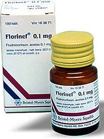 Fludrocortisone is a synthetic corticosteroid that possesses moderate glucocorticoid activity as well as having marked mineralocorticoid potency. By comparison, fludrocortisone has 10 times the glucocorticoid activity and 125 times the mineralocorticoid activity of cortisol. In this regard, fludrocortisone is very different than DOCP, which possess no glucocorticoid activity.
If fludrocortisone acetate is employed as mineralocorticoid supplementation, we recommend an initial oral dosage of approximately 0.02 mg/kg/day.
After initiation of fludrocortisone therapy, serum electrolyte and creatinine concentration should be monitored weekly, with the dosage adjusted by 0.05-0.1 mg/day increments until values have stabilized within the reference range. Once this is achieved, the dogs should be reevaluated monthly for the first 3 to 6 months of therapy, then every 3 to 6 months thereafter.
For dogs that have atypical or secondary Addison’s, mineralocorticoid replacement therapy with DOCP or fludrocortisone aren't needed because the production of aldosterone isn’t effected and the serum electrolytes remain in balance.
Glucocorticoid treatment
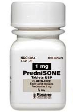
In addition to replacing deficient mineralocorticoids in dogs with Addison's disease, the missing glucocorticoids must also be replaced. This is typically done with an oral form of the synthetic glucocorticoids prednisone, prednisolone, or hydrocortisone. With atypical Addison's and secondary forms of hypoadrenocorticism, glucocorticoid replacement is all that is needed, at least initially, since these dogs do not have serum electrolyte abnormalities.
 |
 |
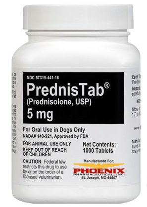 |
The correct dose of the supplemented glucocorticoids, such as prednisone, cannot be measured with a blood test. Rather it's determined by your observations: the lowest dose that keeps your dog symptom free, happy and eating! The most common side effect of overdosage is an increase in thirst and urination, which can be intense in some dogs.
Prognosis of Addison's disease
With proper treatment, the long-term prognosis is excellent. While your dog with Addison’s disease will need medications and monitoring for the rest of his life, most dogs with Addison’s can return to their favorite activities. You will help your dog lead a normal, active and fun-filled life.
Źródło: animalendocrine.blogspot.com
I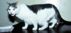 n the past four years, diabetes rates among dogs in the U.S. increased roughly 33% among dogs and 16% among the nation's cat population, per a national analysis of pet health.
According to the report, other common health problems identified among pets include flea and tick infestations, internal parasites, and outer ear inflammations.
See the complete article published today in The Record (Hackensack, New Jersey)
Źródło: animalendocrine.blogspot.com
University of Georgia researchers, studying more than 75,000 dogs from 82 breeds, have found that the causes of canine deaths vary by breed as well as age (1). The study revealed that congenital diseases, trauma and infection are the most frequent causes of deaths in dogs under 2 years old and that cancer risk peaks at about age 10.
The researchers also determined that bigger dog breeds are more vulnerable to musculoskeletal disease, gastrointestinal disease, and cancer. Smaller breed dogs, on the other hand, are more at risk for endocrine and metabolic diseases such as diabetes mellitus and Cushing's disease(1).

Rosie, an 11-year old Bichone Frise with both diabetes and Cushing's disease. The photo on the left is before treatment, whereas the photo on the right is after successful treatment of the Cushing's disease and diabetes.
It has long been recognized that there are patterns in the causes of death for our dogs. This study helps owners know what sort of problems to watch out for in their pets. It helps veterinarians focus on the most likely cause of a particular dog’s illness. And most importantly it guides us in identifying specific risks for individual patients and taking action to minimize these and prevent or delay illness and death.
1. Fleming JM, Creevy KE, Promislow DEL. Mortality in North American Dogs from 1984 to 2004: An Investigation into Age-, Size-, and Breed-Related Causes of Death. Journal of Veterinary Internal Medicine 2011; 25: 187-198.
Źródło: animalendocrine.blogspot.com
 High levels of toxic flame-retardant chemicals including polybrominated diphenyl ethers (PBDE's) were measured in the blood of 18 pet dogs. The measured concentrations of these toxic chemicals were up to 10 times higher as those generally found in humans, according to research published in the journal Environmental Science and Technology (1).
The PBDE compounds are widely used as flame retardants in household furniture and electronics equipment; these chemicals are known to migrate out of the products and enter the environment.
Researchers suggest that pets may act as "biosentinels" for humans who live with them in the same home. "Even though they've been around for quite awhile, we don't know too much about these compounds' toxicological effects on humans or animals," said research scientist Marta Venier (2).
A previous study from the same laboratory showed that pet cats also had much higher serum levels of flame retardants compared to humans, despite sharing the same household environment (3). It has been postulated that these high PBDE levels may contribute to the development of thyroid tumors and hyperthyroidism in cats.
Dogs, in contrast to cats, could be expected to have lower serum levels of flame retardants because they are metabolically better equipped to degrade these compounds. Thus, dogs might be more similar to humans in their response to these environmental chemicals and be better indicators of human exposures to these contaminants.Thus, it was surprising to find the high levels of these toxic compounds in these dog blood samples.
References:
Venier M, Hites RA. Flame retardants in the serum of pet dogs and in their food. Environ Sci Technol. 2011 Article ASAP DOI:10.1021/es1043529
Science News. Toxic Chemicals Found in Pet Dogs.
Dye JA, Venier M, Zhu L, Ward CR, Hites RA, Birnbaum LS. Elevated PBDE levels in pet cats: sentinels for humans? Environ Sci Technol. 2007;41:6350-6356.
Źródło: animalendocrine.blogspot.com
 Hypoadrenocorticism, also called adrenal insufficiency or Addison’s disease, is a disorder in which the adrenal gland does not produce sufficient adrenal hormones that are essential for life. When the adrenal glands fail, the consequences are very severe. Untreated, hypoadrenocorticism may lead to death.
Fortunately, naturally occurring hypoadrenocorticism is extremely rare in cats, with less than 25 cases reported. However, the disease is generally missed because veterinarians rarely consider this problem as a differential diagnosis in cats.
A much more common reason that cats develop hypoadrenocorticism is long-term treatment with steroids for a variety of dermatologic or behavioral reasons. These cats do not show signs of adrenal insufficiency unless the mediation is stopped too abruptly. This is also known as iatrogenic hypoadrenocorticism (see, "What causes this disease in cats," below).
What are the missing hormones in cats with hypoadrenocorticism?
Like dogs or people with Addison’s disease, cats with hypoadrenocorticism are unable to produce one or two steroid hormones, both secreted from the adrenal cortex (outer layer of the adrenal gland).
- The first hormone that's missing is cortisol, which is very important in maintaining a normal metabolism, as well as a general sense of well-being.
- The second hormone that's missing is aldosterone, which manages the water balance and serum electrolytes in the body.
What are the different forms of hypoadrenocorticism in cats?
Hypoadrenocorticism can be divided into primary and secondary subtypes:
- With primary hypoadrenocorticism (Addison's disease), the problem lies in the adrenal gland itself, with atrophy or destruction of the gland.
- With secondary hypoadrenocorticism, the adrenal glands are normal, and the problem lies in the pituitary gland. The pituitary gland normally secretes a hormone called ACTH (adrenocorticotropic hormone) that stimulates the adrenal gland to secrete its hormones. In secondary hypoadrenocorticism, ACTH is not secreted in needed amounts, leading to the secondary adrenal insufficiency.
What causes this disease in cats?
In most cats with naturally occurring hypoadrenocorticism, the exact cause for the adrenal failure is never known for certain.
Many cats, however, develop the problem because of iatrogenic causes. The term iatrogenic means that the disease is caused by, or results from, a medical or surgical treatment for another problem (i.e., the disease did not develop spontaneously). In cats with primary hypoadrenocorticism, the following causes must be considered.
- Most cats that develop Addison’s disease spontaneously appear to have an autoimmune condition in which the body destroys part of the adrenal cortex.
- Infiltrative conditions such as lymphoma (a form of cancer) can destroy the adrenal gland to cause Addison’s disease.
- Surgical removal of both adrenal glands for treatment of feline Cushing’s disease will result in iatrogenic Addison’s disease, but this is a rarely used operation for cats.
- Rarely, primary adrenal failure can occur secondary to trauma. This may be temporary or permanent.
In cats with secondary hypoadrenocorticism, the following underlying causes should be excluded. Again, all of these conditions are associated with deficient secretion of the pituitary hormone, ACTH.
Congenital deficiency of ACTH secretion (not yet reported in cats).
Pituitary tumors, inflammation, or trauma that have destroyed most of the ACTH-secreting cells in the pituitary gland, leading to deficient ACTH secretion.
Iatrogenic hypoadrenocorticism that results from administration of large doses of cortisone-like hormones (glucocorticoids or progesterone-like drugs), which are then withdrawn too rapidly. In this case, the term iatrogenic means that the hypoadrenocorticism is caused by the high-dose glucocorticoid or progestin (megestrol acetate; trade names, Ovaban or Megace) treatment.
Chain of events leading to severe hypoadrenocorticism in cats
With primary hypoadrenocorticism (Addison's disease), cats develop complete adrenocortical destruction with both cortisol and aldosterone deficiencies.
Aldosterone is the main mineralocorticoid hormone, and it affects the levels of potassium, sodium, and chloride in the blood. Low levels of aldosterone cause potassium to gradually build up in the blood and, in severe cases, cause the heart to slow down or beat irregularly. Some cats have such a slow heart rate (50 beats per minute or lower) that they can become weak or go into shock.
More commonly, cats will develop iatrogenic, secondary hypoadrenocorticism (see above, What causes this disease in cats) to treatment with steroid-like drugs used for anti-inflammatory purposes. This subgroup of hypoadrenal cats is only deficient in cortisol and maintains a normal aldosterone level. These cats are more difficult to diagnose because their signs are milder and the serum potassium, sodium, and chloride values all remain normal.
What are the clinical signs and symptoms of feline hypoadrenocorticism?
Signs of hypoadrenocorticism may include repeated episodes of vomiting and diarrhea, loss of appetite, dehydration, and gradual, but severe, weight loss.
Because the clinical signs of hypoadrenocorticism are vague and nonspecific, it can be difficult to diagnose in the earlier stages of disease. Therefore, severe consequences, such as shock and evidence of kidney failure, can develop suddenly in some cats with complete adrenocortical destruction.
In cats with secondary hypoadrenocorticism, the clinical signs generally are not as severe and are usually not life threatening.
 How is feline hypoadrenocorticism diagnosed?
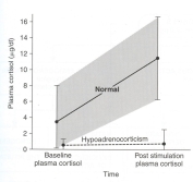
A veterinarian may suspect hypoadrenocorticism based on the cat’s history (including past treatment with steroids or progestins), clinical signs, and certain laboratory abnormalities, such as low serum sodium and high potassium concentrations. However, one must specifically evaluate adrenal function to document low cortisol levels to definitively diagnose hypoadrenocorticism.
The diagnosis of hypoadrenocorticism is confirmed by lack of cortisol response to ACTH administration. It is important to recognize that the reference range for the post ACTH cortisol is lower in cats than in dogs. To confirm a diagnosis of hypoadrenocorticism, however, the serum concentrations of both the basal cortisol level and ACTH-stimulated cortisol levels are low (generally less than 2 µg/dl).
 How is primary hypoadrenocorticism (Addison’s disease) treated?
Untreated, primary hypoadrenocorticism (Addison’s disease) can lead to an adrenal crisis. An adrenal crisis is a medical emergency that requires intravenous fluids to restore the body’s levels of fluids, salts, and sugar to normal. Once stabilized, the cat can then be treated with hormone replacement therapy (either orally or by injection). With proper treatment, the long-term prognosis is excellent.
Long-tem treatment of primary hypoadrenocorticism (Addison’s disease) includes treatment with either both missing adrenal hormones. For mineralocorticoid replacement, either oral fludrocortisone (trade name, Florinef) or injectable desoxycortisone privalate (DOCP, trade name, Percorten) are given. In cats, glucocorticoid replacement is best given as oral or injectable prednisolone.
Response to treatment is similar although clinical signs such as anorexia, lethargy, and weakness may sometimes take longer to resolve than in dogs. Prognosis for long-term survival is good with the exception of cats in which the underlying cause is adrenal neoplasia.
How is iatrogenic hypoadrenocorticism (secondary to overtreatment with steroids or progestins) treated?
In cats with iatrogenic hypoadrenocorticism due to overtreatment with steroids for nonendocrine problems, treatment simply involves reinstituting glucocorticoid replacement at a gradual tapering dosage over weeks to months. This allows the pituitary ACTH-secreting cells to recover and stimulate normal cortisol secretion once again.
Źródło: animalendocrine.blogspot.com
 The Animal Endocrine Clinic recently partnered with Anjellicle Cats Rescue and Ready for Rescue animal shelters to provide radioactive iodine treatment to two hyperthyroid cats. The success of the advanced medical treatment, pioneered by Dr. Peterson, will allow the cats a chance at being adopted into new homes.
Molly, a 15 year old cat was removed from the Animal Care & Control of NYC’s “kill list” by the Anjellicle Cats Rescue. Despite the fact that Molly had become very thin and had the typical increased appetite, common to hyperthyroid cats, the rescue felt that they could find her a home. A “foster mother” with the shelter, Margaret Cozzo had heard of Dr. Peterson’s Animal Endocrine Clinic and reached out to him for help with the cat’s disease.
Said the Director of Anjellicle Cats Rescue, Kathryn Willis, “We are very pleased with the success of the treatment that Molly received at the Animal Endocrine Clinic. She has regained the weight and the spirit that she had lost while sick. She has a great disposition and will make a lovely pet for anyone.”
Foxy, a nine year old Tabby, was another cat that was saved from the “kill list” by the organization, Ready for Rescue. Foxy was also small and underweight as a result of her hyperthyroid condition. Initially treated with the daily medication, Methimazole, she experienced bad side effects. Looking for a better option, her “foster mother,” volunteer Karin Felix-Faure, had lived near the Animal Endocrine Clinic’s Manhattan location and sought out Dr. Peterson’s expertise.
Dr. Mark Peterson treated both cats at the Animal Endocrine Clinic using the most advanced medical diagnostics and treatments available for feline hyperthyroidism. Using nuclear imaging, he confirmed that both cats were hyperthyroid and then treated them with radioiodine treatment. Donations and discounts from the AEC helped pay for the cats’ treatments.
“We almost lost Foxy because she couldn’t handle the daily medication,” said Doug Halsey of Ready for Rescue. “If it hadn’t been for Dr. Peterson’s advanced treatment, Foxy would not be alive today. How cruel it would have been for her to have been removed from the ‘kill list’ and then die while receiving treatment.”
Dr. Peterson offered, “Radioactive iodine treatment is the home humane and efficient way to cure Hyperthyroidism, a common disease in cats. If left untreated, it can lead to death. We were thrilled to be able to provide these cats with a second chance at being loved and cared for in new homes. One subcutaneous treatment was all that was needed to cure both cats.”
About Anjellicle Cats Rescue:
Anjellicle Cats Rescue is an all-volunteer, not-for-profit 501(c)(3) organization. The rescue is also a member of the Mayor’s Alliance for NYC’s Animals and a New Hope Partner with the New York Animal Care & Control (ACC). The rescue has a no-kill policy and holds adoption events throughout New York City and in Connecticut. For more information about Anjellicle Cats Rescue visit http://www.anjelliclecats.com/
About Ready for Rescue:
Ready for Rescue is a New York City-based animal rescue group dedicated to saving at risk animals with a focus on cats and dogs who are suffering in the New York City shelter system. The shelter shows cats and dogs up for adoption at The Pet Health Store in Manhattan on Sundays, Noon – 4pm. For more information about Ready for Rescue visit http://ready4rescue.org/ About The Animal Endocrine Clinic: The Animal Endocrine Clinic (AEC) is a specialized veterinary practice that diagnoses and treats cats and dogs with endocrine (hormonal) disorders. AEC has three divisions:
- Hypurrcat exclusively treats hyperthyroid cats with radioactive iodine;
- The AEC's Endocrine Clinic is dedicated to diagnosing and treating dogs and cats with endocrine disorders, such as diabetes or Cushing’s disease; and
- Nuclear Imaging for Animals is a state-of-the-art medical imaging facility that performs nuclear scanning to diagnose thyroid, bone, liver, and kidney diseases in dogs and cats.
AEC has clinics in Manhattan and Westchester County and treats clients in New York City, Long Island, Westchester County, New Jersey and Connecticut. It is the only practice of its kind in the United States. For more information about AEC visit www.animalendocrine.com. Link to Press Release:
http://www.pr.com/press-release/317450
Źródło: animalendocrine.blogspot.com
 Bailey is a 14-year-old male neutered domestic short hair who was diagnosed with hyperthyroidism by another veterinarian about 3 years ago and has been on methimazole since then. He also has a history of chronic constipation that has been well controlled in the last year on enulose (Lactulose) only.
I last saw Bailey a year ago, at which point he was doing well clinically but had lost about 1 lb. since his last check. At that time, he was on 5 mg of methimazole BID. A geriatric profile at that time showed a total T4 of 3.3 µg/dl, a normal CBC and serum chemistry profile (creatinine = 1.5 mg/dl, BUN = 23 mg/dl), and a urine specific gravity of 1.065. I recommended increasing his dose of methimazole to 7.5 mg in the morning and 5 mg in the evening and rechecking in 4-6 weeks.
The owner did increase the daily methimazole dose but did not follow up with us until now. Bailey has lost an additional 3 lbs. since his last exam and has developed polyphagia, polyuria, and polydipsia over the last month.
On exam yesterday, the cat now has a fairly large mass in his left ventral neck area (about 3-4 cm, slightly firm and irregular), which I had not noted on the last exam. His blood work from yesterday showed a T4 of 1.9 µg/dl, and very normal renal function (serum creatinine = 1.1 mg/dl; BUN = 22 mg/dl, USG =1.030).
So, I'm suspicious that the thyroid mass is or has become a carcinoma at this point. But would you expect the cat to lose weight secondary to a thyroid carcinoma even if the T4 is within normal range? Could it be secreting another hormone to cause the weight loss?
I thought I should recommend full-body radiographs to try to rule out any metastasis or other obvious neoplasia, and then consider surgical removal and biopsy of the mass. I've read that it can be helpful to follow-up with I-131 treatment after excision of a thyroid carcinoma, so I would speak to the owner about referral for that.
Does that sound like a reasonable course of action to you or are there other things you would recommend doing first? Thank you very much for any help or advice you can give me on this perplexing case.
My Response:
There are many ways you could go in your workup of this cat. Most cats that I see who are loosing weight while on treatment with methimazole have high serum T4 concentrations, which can explain the weight loss. Obviously, that's not the case here, which makes this cat more interesting!
Many cats (in fact, probably nearly all hyperthyroid cats) will have an increase in goiter size with time — that makes sense since we aren't inhibiting thyroid tumor growth with the methimazole. We are only blocking thyroid hormone secretion with the drug.
Recently, a paper was published showing that on thyroid biopsy, some cats with long-standing hyperthyroidism had evidence of transformation of thyroid adenoma to carcinoma (1). I do believe that this happens more than we realize, and I now see almost a cat a month with thyroid carcinoma. Almost all cats with thyroid carcinoma that I see have been on methimazole for longer than 2 years.
So in your cat, the enlargement of the thyroid mass could indicate that the tumor has simply grown larger with time, or it could indicate malignant transformation. As you indicated, thyroid biopsy would be helpful in making that diagnosis. Many of these cats have extension or metastasis into the thoracic cavity so you might not be able to cure that cat with surgical thyroidectomy if that is the case. What I like to do in that situation is to perform a thyroid scan (thyroid scintigraphy) prior to surgery, which we certainly could do in this cat. This will tell us where the thyroid tumor tissue is located and help direct what needs to be removed or biopsied, if the cat does go to surgery.
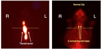
Thyroid Scan of cat with large thyroid carcinoma (left) compared to normal cat (right)
Notice the extension & invasion of the thyroid tumor into the thoracic cavity
(horizontal line indicates the region of the thoracic inlet)
That all said, why is your cat loosing weight despite a normal serum T4? With the polyphagia, the cat should bee taking in enough calories. If there vomiting or diarrhea? (I assume that diarrhea isn't present because of the constipation.)
Cats with weight loss that are eating normally must have either increased loss of glucose in the urine or impaired absorption of nutrients from the GIT. To that end, I would recommend that you perform an abdominal ultrasound, in addition to your full-body x-rays, prior to either thyroid biopsy or thyroid scintigraphy. Urine culture should also be considered to exclude pyelonephritis.
As far as other hormones being secreted, I'd suggest that you also measure a serum free T4 and T3 concentration on this cat. It's possible that the free T4 or T3 concentrations are still high and that could explain some of weight loss in this cat.
Reference:
1. Hibbert A, Gruffydd-Jones T, Barrett EL, Day MJ, Harvey AM. Feline thyroid carcinoma: diagnosis and response to high-dose radioactive iodine treatment. J Feline Med Surg. 2009 11:116-24.
Źródło: endocrinevet.blogspot.com

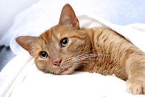
Rosie came to us roughly a year ago exhibiting all of the classic signs of Cushing's disease, which I covered in my last blog post:
increased thirst and urination
increased appetite
excessive panting (see photo on left)
lethargy
pot belly appearance (see photo on left)
weight gain (see photo on left)
hair loss: hair thinning on trunk, bald tail (see photo on left)
I diagnosed her as having Cushing's disease and prescribed a regimen of trilostane (Vetoryl), given at a dosage of 30 mg twice a day.
The photo on the right was taken at her one-year check up. As you can see from the photos, she's lost weight (8 pounds) as well as her pot belly. She is much more active and no longer excessively thirsty or panting.
Źródło: animalendocrine.blogspot.com
Over the past week I have received a number of questions about radiation both from pet owners and veterinarians about the danger from the nuclear fallout from Japan, not for their human families but also for their dogs and cats.
 The question is: should we all be buying potassium iodide for ourselves? Should we also give this to our pets? Since dogs and cats are smaller than we are, could they be impacted by smaller amounts of radiation? What should we do?
The answer to these questions, at least for the time being, is not to take potassium iodide or administer the drug to our pets. So far, the amount of radiation coming here from Japan has been termed negligible by our government.
What's more, potassium iodide only helps pets (or people) to deal with radioactive particles (I-131) which ultimately impact the thyroid gland, not other organs or illnesses which may result from excessive exposure to radiation. I understand that we care about our pets and want to be proactive, but we might do more harm than good by giving this iodide supplement.
 How does potassium iodine work?
The thyroid gland is the only tissue in the body that wants or needs iodine to perform its normal function (i.e., to make the thyroid hormones T4 and T3, both of which contain iodine in their hormone structure). As you can see, in the figure on the right, thyroxine or T4 (the main thyroid hormone secreted by the thyroid gland), contains 4 iodine groups on the hormone molecule. T3, the other main thyroid hormone, contains 3 iodine groups on the molecule.
By saturating the thyroid with stable iodide (i.e., non-radioactive iodine), potassium iodide works to block the effects of radioiodine (I-131) on the thyroid gland. In other words, if your thyroid already has all the iodine it needs it will refuse to take up the I-131.
Of course, there's a catch. Doses of potassium iodide high enough to protect your thyroid from I-131 have some nasty side effects.
What are the adverse effects of potassium iodide?
Side effects associated with administration of potassium iodide may include gastrointestinal upset, allergic reactions, skin rashes, salivary gland inflammation, hyperthyroidism, or hypothyroidism. That is a pretty impressive list of side effects!
So what should we do?
Since radioactive iodine (I-131) decays rapidly, current estimates indicate there will not be a hazardous level of radiation reaching the United States from this accident. If and when an exposure does warrant the use of potassium iodide, it should be taken as directed by physicians or public health authorities until the risk for significant exposure to radioactive iodine dissipates, but probably for no more than 1 to 2 weeks.
Again, unless the experts declare that radiation levels are high enough, we shouldn't take it or administer it to our pets. If you're determined to purchase potassium iodide for you or your pet, please beware —many legitimate outlets are sold out, particularly if you purchase the potassium iodide online.
Źródło: animalendocrine.blogspot.com
This week, one of my veterinary colleagues returned from a 3-week visit to Japan. She was obviously worried about possible radiation exposure in the aftermath of the Japan nuclear crisis and asked for my advice about how she could check for radiation contamination. I asked her to come to my office today so that I could monitor her for any radiation contamination. I have the radiation instruments in both of my offices because we routinely treat hyperthyroid cats with radioactive iodine (I-131). (See my website for more information.) My staff and I use this radiation detection equipment on a daily basis in order to check for contamination, both on our bodies and well as in our work environment (this is a safety precaution - we do NOT expect or plan on becoming contaminated!).
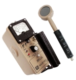
I first monitored by colleague by use of a general purpose survey meter (Geiger counter). With this meter, I've attached a GM (geiger-mueller) pancake detector, which is sensitive to alpha, beta and gamma radiation and is the industry standard for detecting contamination.
Fortunately, I found absolutely no detectable levels of external contamination with my measurements.

Dr. Peterson checking for external radiation contamination
We next measured my colleagues thyroid gland for internal contamination. As you may know, the principal radiation source of concern with the nuclear reactor accident in Japan is the release of radioactive iodine (I-131). This radioactive isotope that presents a special risk to health because iodine is normally concentrated in the thyroid gland. Exposure of the thyroid to high levels of radioactive iodine may lead to development of thyroid nodules and thyroid cancer years later.

Counting thyroid gland for I-131 contamination
To measure for internal (thyroid) contamination of I-131, we used a general purpose radiation scaler/ratemeter attached to a shielded well gamma radiation counter. This is a very sensitive instrument, which allows us to detect very tiny amounts of gamma radiation, including the gamma rays (photons) emitted from I-131.

Radiation counts below background readings, indicating no contamination
Fortunately, the thyroid counts measured in my college (373 counts per minute or cpm) were below the background radiation counts of 385 cpm. This demonstrated that there was NO internal thyroid radiation. Overall, we found NO external or internal contamination in my colleague. All good news! The fact that we measured a background radiation count is a normal, expected finding: background radiation is constantly present in the environment and is emitted from a variety of natural and artificial sources. See this article for more information on background radiation.
Źródło: animalendocrine.blogspot.com