I have a 7-year old female spayed Siamese cat that has been on Hill's y/d food for the past 2 months. The cat's thyroid levels have not come down (still > 15 μg/dl) and she is miserable on the food. She acts like she's starving (possibly from the uncontrolled thyroid) and is trying to steal food from the owner.

I am discontinuing the food but was wondering if I need to wait a certain amount of time before starting her back on methimazole?

My Response:

I've heard the same story from many cat owners and veterinarians. The y/d just doesn't appear to look and taste very appetizing to many cats (the food certainly looks pretty gross to me, but I must admit — I haven't tasted it!).

If the cat's serum T4 value is still high now, I'd switch the cat to a good diet (higher in protein, lower in carbs) that she wants to eat and start methimazole now.
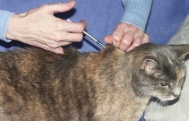
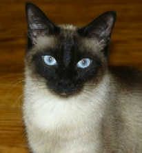
  • A 7-year old Siamese could live a very long time, possibly another 15 years! I'd talk to the owners about the following facts when you discuss how to treat the cat's hyperthyroidism (1-5): 
  • This cat, like all hyperthyroid cats, has a thyroid tumor
  • The thyroid tumor and hyperthyroidism will never go into spontaneous remission
  • The thyroid tumor will continue to grow larger with time
  • In some cats treated long term medically, the benign tumor will transform into a malignant thyroid carcinoma. In my studies, the incidence of thyroid carcinoma is above 20% in cats treated medically for longer than 4 years (6).
The bottom line:
If this was my own cat, I'd either do surgery or radioiodine to cure the cat's hyperthyroidism rather than trying to manage it with an iodine deficient diet (Hill's y/d) or methimazole for the rest of her life (1-5,7,8). She is already suffering from a severe case of hyperthyroidism, which will only worsen with time.

In the long run, a definitive treatment would be the wisest (and most cost effective) means of treating this relatively young cat.

References & Suggested Reading:
  1. Baral R, Peterson ME. Thyroid gland disorders. In: Little, S.E. (ed), The Cat: Clinical Medicine and Management. Philadelphia, Elsevier Saunders 2012; 571-592. 
  2. Peterson ME: Hyperthyroidism, In: Ettinger SJ, Feldman EC (eds): Textbook of Veterinary Internal Medicine: Diseases of the Dog and Cat (Fifth Edition). Philadelphia, WB Saunders Co. 2000; pp 1400-1419.
  3. Peterson ME: Hyperthyroidism in cats. In: Melian C (ed): Manual de Endocrinología en Pequeños Animales (Manual of Small Animal Endocrinology). Multimedica, Barcelona, Spain, 2008, pp 127-168.
  4. Mooney CT, Peterson ME: Feline hyperthyroidism, In: Mooney C.T., Peterson M.E. (eds), Manual of Canine and Feline Endocrinology (Fourth Ed), Quedgeley, Gloucester, British Small Animal Veterinary Association, 2012; in press. 
  5. Peterson ME: Hyperthyroidism in cats, In: Rand, J (ed), Clinical Endocrinology of Companion Animals. New York, Wiley-Blackwell, 2012; in press.
  6. Peterson ME, Broome MR. Thyroid scintigraphic findings in 917 cats with hyperthryoidism. Journal of Veterinary Internal Medicine 2012; in press.
  7. Peterson ME: Radioiodine treatment for hyperthyroidism. Clinical Techniques in Small Animal Practice 2006;21:34-39.
  8. Peterson ME: Radioiodine for hyperthyroidism. In: Bonagura JD, Twedt DC (eds): Current Veterinary Therapy XIV. Philadelphia, Saunders Elsevier, 2008, pp 180-184.

Źródło: endocrinevet.blogspot.com

How long human, recombinant NPH insulin can be used once the vial is opened? 

Our local pharmacist told one of my diabetic dog owners that the human NPH insulin she is using for her dog would only be effective for 30 days. After that time, the pharmacist claims the vial should be discarded and replaced with a fresh vial of insulin.  

The pharmacy also told the owner that the insulin should not be refrigerated.

I'm confused. I always thought that NPH insulin should be refrigerated and is good 'till the last drop!"

My Response

The pharmacist is right and wrong in his advise — it just "depends."

In general, open NPH insulin vials in human diabetic patients are kept unrefrigerated to minimize local injection site irritation, which may occur after injection of cold insulin solutions.

Human patients are also told to replace the insulin with a new vial every 4 to 6 weeks. Why? Because the insulin my lose some of its potency after the vial has been in use for >30 days when stored at room temperature.

In both humans and animals, we can recommend general guidelines for the storage and handling of NPH insulin preparations, which include Humulin N, Novolin N, or Humulin/ReliOn NPH (1).

  • All opened insulin vials should be inspected daily for physical changes, such as clumping, frosting, precipitation, or discoloration, that may be accompanied by a loss of potency. 
  • Insulin vials should be optimally stored at refrigerated temperatures (36–46°F).
  • The insulin vial should never be allowed to freeze.
  • Insulin vials should never be used after the expiration date printed on the label and carton.
  • Unrefrigerated insulin vials that are in use should be kept cool and away from excess heat or sunlight.

We ask our pet owners to refrigerate the insulin, which tends to extend the shelf life of the insulin.
Many insulins have good potency for 6 months; however, contamination from multiple sticks can be an issue, so many veterinarians recommend switching every 3 months or so.

Whenever human insulin vials are stored under refrigeration and are used beyond 30 days, the stability of these vials may be affected by a number of factors, including:


  • the number of injections per day
  • the volume of insulin remaining in the vial
  • exposure to light,
  • degree of agitation used for dose preparation 

The impact of such factors is difficult to measure, so veterinarians should not be reluctant to replace an insulin vial with a fresh one if diabetes regulation due to loss of insulin potency becomes a concern.

But you are correct: in many diabetic dogs, the NPH insulin is indeed good "till the last drop" is given.

Reference


  1. Grajower MM, Fraser CG, Holcombe JH, et al. How long should insulin be used once a vial is started? Diabetes Care. 2003;26:2665-2666.


Źródło: endocrinevet.blogspot.com

Ultrasound revealing pancreatic cyst
in a 10-year old diabetic
My patient is a 10-year old male neutered DSH that was treated for several years for diabetes mellitus. He went in to remission and was doing well until this summer. He was presented after having a "weird" episode after being in a room with an undiagnosed "toxin."

Physical examination revealed a palpable abdominal fluid wave and mild muscle wasting in the hindend. Blood work at that time revealed a mild evaluation in globulins but no other significant changes. 

An abdominal ultrasound revealed a hypoechoic pancreas with a large pancreatic cyst. No other abnormalities were noted. The effusion had an elevated total protein and cytology revealed very few cells with one macrophage that was suspicious for corona virus on IFA. A feline pancreatic lipase immunoreactivity (fPLI) concentration was elevated at 36.

He was treated with supportive care and improved over the next few weeks, the ascites resolved but the pancreatic cyst remains present. He was noted to have a ravenous appetite but no other clinical signs. a recheck fPLI had decreased to 6.5, a T4 was within normal limits, as was a serum trypin-like immunoreactivity (TLI) concentration. He has put more weight on after the owner increased his food ration. Over the past few weeks he has been doing well but the owner notes that he is licking smacking excessively and has loss some hair on the ventral tail and over the tarsus. We would appreciate your input on the case and your thoughts on aspirating the cyst.

Thank you in advance for your time and expertise.

My Response:

A pancreatic cyst is a rare finding in cats (1-4). In most cats, they are a benign incidental finding. the cysts can be associated with pancreatitis. It has been hypothesized that diabetes mellitus may develop secondary to a pancreatic cyst, but this does not appear to be the case with your cat inasmuch as the diabetes had been present for several years prior to the detection of the cyst.

In general, the treatment approach to a pancreatic cyst or pseudocyst involves everything from benign neglect if the cyst is small or the cat is asymtomatic, draining the cyst using an ultrasound guided procedure, or surgical removal, especially in cases where the cyst is large and quickly refills with fluid after drainage.

References:
  1. Coleman MG, Robson MC, Harvey C.  Pancreatic cyst in a cat. N Z Vet J. 2005;53:157-159.
  2. Branter EM, Viviano KR.  Multiple recurrent pancreatic cysts with associated pancreatic inflammation and atrophy in a cat. J Feline Med Surg. 2010;12:822-827.
  3. VanEnkevort BA, O'Brien RT, Young KM. Pancreatic pseudocysts in 4 dogs and 2 cats: ultrasonographic and clinicopathologic findings. J Vet Intern Med. 1999;13:309-313.
  4. Hines BL, Salisbury SK, Jakovljevic S, et al. Pancreatic pseudocyst associated with chronic-active necrotizing pancreatitis in a cat.  J Am Anim Hosp Assoc 1996;32:147-152.


Źródło: endocrinevet.blogspot.com


I had attended your endocrine lectures at the annual meeting of the Minnesota Veterinary Medical Association last month and heard your great talk on how you workup and treat dogs with Cushing's syndrome.

My patient is a 9-year old, FS, Brittany Spaniel, weighing about 20 kg. She has a 6-month history of increased drinking, urination, hunger, and weight gain. 

On her routine blood tests, she had high serum alanine aminotransferase activity (ALT, 152 U/L; reference range, 12-118 U/L), high serum alkaline phosphatase activity (1998 U/L; reference range, 23-212 U/L). The serum cholesterol was also elevated  at 459 mg/dl (reference range, 110-320 mg/dl). I did a serum bile acids test, which was normal.

After I heard your lecture, I did a low-dose dexamethasone suppression test, with the following cortisol results. 

  • Cortisol basal sample: 3.3 μg/dl (reference range, 1-5 μg/d)
  • Cortisol 4 hr: 2.2 μg/dl (reference range, 0.0-1.4 μg/d)
  • Cortisol 8 hr: 3.5 μg/dl (reference range, 0.0-1.4 μg/d)

I know that the lack of cortisol suppression is diagnostic for Cushing's syndrome but the results do not differentiate between pituitary-dependent hyperadrenocorticism and functional adrenal tumor.

The owner is interested in doing an abdominal ultrasound and wants to pursue treatment eventually. So we are planning to set up an appointment for an ultrasound as the next step, but I did have some questions in terms of longer-term outcome for dogs with Cushing's disease. With some of our other suspected cases, the owners have not pursued pituitary-adrenal function testing or treatment due to cost, so I haven't actually had any that I've gone on to treat (I've also only been practicing since July 2010).  


  1. If it is an adrenal tumor, you had recommend treating with trilostane for several weeks prior to surgical adrenalectomy, correct? Does this help to minimize post-operative complications?
  2. And if they go on to surgery, and it comes back as benign, do most dogs do pretty well after long-term?
  3. If the owner cannot afford/does not want to pursue surgery, can they do relatively well on trilostane (provided it isn't a carcinoma)?
  4. If pituitary-dependent and treated with trilostane, do dogs do pretty well long-term? I wasn't sure about their long-term prognosis.

Thanks for you time in advance. I really appreciate any insight you're able to give me since again, we haven't really treated many dogs at my clinic.

My Response:

In answer to your questions:


  1. Yes, if this dog has an adrenal tumor, I recommend treating with trilostane (Vetoryl, Dechra Animal Health) for 4-6 weeks prior to surgery (1,2). Controlling the high cortisol values prior to surgery helps to minimize post-operative complications, such as thromboembolism, poor wound healing, and infections.
  2. If the dog has a benign adrenal tumor which is removed surgically, the dog would be cured (1). Post-operatively, she will go through a period of adrenal insufficiency (remember that the contralateral adrenal cortex is suppressed and will take a few weeks to start working again). During the immediate post-operative period, glucocorticoid supplementation should be initiated. The doses of the daily glucocorticoids should then be slowly tapered over the next 6-8 weeks, when they can generally be discontinued in most dogs.
  3. If the owner cannot afford the cost of adrenalectomy or does not want to pursue surgery, most dogs can be controlled relatively well on trilostane (Vetoryl) but will never be cured (1-3). Overall, the prognosis with medical treatment is not as good as surgical treatment. If the dog has adrenal carcinoma, medical treatment will not prevent tumor growth, invasion into the vena cava, or metastasis (1).
  4. If the dog has pituitary-dependent hyperadrenocorticism (PDH) and is treated with trilostane (Vetoryl), most dogs do well when treated with either mitotane (Lysodren) or trilostane. Studies have reported that there is no difference in survival between use of either of these drugs (4,5).

Botton line— We need a differentiating test to help guide our treatment recommendations and determine prognosis. Let me know when you have the results of the abdominal ultrasound on this dog.

Follow-up:

The abdominal ultrasound was just done this morning. Both adrenals were at the upper end of normal size —not nodular— and no masses found on either adrenal gland. 

So these imaging results are consistent with pituitary-dependent Cushing's disease, correct?

My Response:

Yes, your dog has PDH and can be treated with either trilostane or mitotane.

Remember to explain to the owner, however, the dog's problem lies in the pituitary gland, not the adrenal glands. Most of these dogs will have small pituitary tumors that tend to grow slowly over time (1,6,7). Others, however, have large pituitary tumors at time of diagnosis and some of these tumors expand more rapidly and can cause compression of the hypothalamus and lead to neurological signs.

Hopefully, your patient will be well-controlled with the medical treatment and will die of old age before the development of a large, invasive pituitary tumor becomes and issue.

References:


  1. Melián C, M. Pérez-Alenza, D, Peterson ME. Hyperadrenocorticism in dogs, In: Ettinger SJ (ed): Textbook of Veterinary Internal Medicine: Diseases of the Dog and Cat (Seventh Edition). Philadelphia, Saunders Elsevier, 2010; pp. 1816-1840.
  2. Ramsey IK. Trilostane in dogs. The Veterinary Clinics of North America Small Animal Practice 2010;40:269-283. 
  3. Helm JR, McLauchlan G, Boden LA, et al. A comparison of factors that influence survival in dogs with adrenal-dependent hyperadrenocorticism treated with mitotane or trilostane. Journal of Veterinary Internal Medicine 2011; 25:251-260.  
  4. Clemente M, De Andrés PJ, Arenas C, et al. Comparison of non-selective adrenocorticolysis with mitotane or trilostane for the treatment of dogs with pituitary-dependent hyperadrenocorticism. Veterinary Record 2007;161:805-809.
  5. Kintzer PP, Peterson ME. Mitotane (o,p'-DDD) treatment of 200 dogs with pituitary-dependent hyperadrenocorticism. Journal of Veterinary Internal Medicine  1991;5:182-90. 
  6. Ihle SL. Pituitary corticotroph macrotumors. Diagnosis and treatment. The Veterinary Clinics of North America Small Animal Practice 1997;27:287-297. 
  7. Kent MS, Bommarito D, Feldman E, et al. Survival, neurologic response, and prognostic factors in dogs with pituitary masses treated with radiation therapy and untreated dogs. Journal of Veterinary Internal Medicine 2007;21:1027-1033.

Źródło: endocrinevet.blogspot.com

'm writing about a 7-year-old lab that present today for signs associated with hypoglycemia.He has been progressive lethargic for the last week. The owner noted staggering and weakness and brought him into our clinic for evaluation yesterday.

On examination, the dog was quiet but clinically normal. His blood glucose concentration, however, was low at 26 mg/dl (reference range, 70-125 mg/dl). All other serum chemistry results (including serum sodium and potassium were normal. Radiographs of the chest and abdomen were unremarkable. The owner has been quizzed at length about possible xylitol ingestion, but there is no history of possible ingestion.

We hospitalized the dog for observation and started a 5% dextrose drip overnight. This morning, the blood glucose was still quite low (only 47 mg/dl) after being off dextrose for about an hour. I continued the glucose drip (raised it to a 10% drip), and the blood glucose concentrations ranged from 41 to 70 mg/dl throughout the day. He is eating well and alert on the IV glucose supplementation.

My rule outs are the following intoxications or medical disorders:

- Xylitol ingestion
- Atypical Addison's (the serum electrolytes and Na:K ratio were normal)
- Insulinoma (insulin-secreting pancreatic islet cell tumor)
- Portosystemic shunt

I have submitted an serum insulin level at the time when the dog's blood glucose was very low (42 mg/dl) and the results are still pending. I've also submitted a baseline cortisol concentration to help rule out Addison's disease. I have not measured his serum bile acids because I did not want to fast him just yet.

Any other thoughts, suggestions for diagnostics? Anything I am missing?


My Response:

You have the correct list of differential diagnoses and are working your way through the list properly. I'd agree with not fasting the dog — I'd just do a random bile acid measurement to first see it that's abnormal.

If this dog has an insulinoma, we have to remember that IV administration of dextrose may stimulate secretion by the pancreatic tumor. Insulin release post-hyperglycemia often results in rebound hypoglycemia, which necessitates additional dextrose administration and leads the clinician into a cyclical hyperglycemia/hypoglycemia "chase" which can be difficult to terminate.

In this cases, it an be really helpful to just use glucagon instead. Glucagon for injection (1 mg vial) is reconstituted according to the manufacturer's instructions with supplied diluent, then added to 1000 ml of 0.9% NaCl. The 1000 ng/ml solution is administered intravenously as a constant rate infusion (CRI) with the use of a syringe infusion pump. The glucagon CRI is initially given as a bolus of 50 ng/kg, then administered at a rate of 10 to 15 ng/kg/min. The dose may need to be increased up to 40 ng/kg/min as needed to maintain euglycemia.

Additional Followup:

I've put the dog on a glucagon constant rate infusion and it's been working great to maintain the blood glucose concentration in the normal range in the hospital!

The dog's resting serum cortisol concentration was normal at 3.2 µg/dl, so Addison's disease appears highly unlikely. My serum insulin and bile acid results still pending.

The owner has been internet investigating and he told me that his dog has eaten a birch limb/stick last week. I read that xylitol is derived from hardwoods such as birch — Is there any way the xylan from the birch could be metabolized to xylitol in the dog?


My Response:

You're right, the normal serum cortisol (>2.0 µg/dl) rules out Addison's disease.

As far as xylitol toxicity is concerned, I did some research myself and found that birch wood itself doesn't contain xylitol so that's unlikely to be the cause of the hypoglycemia. And this would be especially true since it was consumed a week ago, and you wouldn't expect continued hypoglycemia if the problem was due to xylitol toxicity.

Outcome:

The dog's serum insulin concentration, collected at the time of severe hypoglycemia, was extremely high (402 pmol/L; reference range, 60-230 pmol/L). The bile acids values were normal. An abdominal ultrasound revealed a pancreatic mass with several hypoechoic areas in liver, consistent with pancreatic neoplasia with metastasis to the liver. A trial of diazoxide was performed at home, but hypoglycemia persisted.

Based on the poor prognosis and lack of response to diazoxide, the owners elected euthanasia. Multiple pancreatic and hepatic nodules were identified at necropsy; histopathology confirmed beta cell islet cell neoplasia with metastasis to the liver.
                                                                                                                                                                   
Final Diagnosis: Insulinoma (insulin-secreting carcinoma of pancreas), producing hypoglycemia.

References:


- Dunayer EK. Hypoglycemia following canine ingestion of xylitol-containing gum. Veterinary and Human Toxicology 2004;46:87-88.
- Leifer CE, Peterson ME, Matus RE. Insulin-secreting tumor: diagnosis and medical and surgical management in 55 dogs. Journal of the American Veterinary Medical Association 1986;188:60-64.
- Smith SA. Miscellaneous Endocrine Disorders. In Morgan RH, Bright, R, and Swartout MS (eds). Handbook of Small Animal Practice, Fourth Edition. W. B. Saunders, Philadelphia, PA. 2003:731-751.
- Fischer JR, Smith SA, Harkin, KR. Glucagon constant rate infusion: a novel strategy for the management of hyperinsulinemic-hypoglycemic crisis in the dog. Journal of the American Animal Hospital Association 2000; 36:27-32.
- Lennon EM, Boyle TE, Hutchins RG, et al: Use of basal serum or plasma cortisol concentrations to rule out a diagnosis of hypoadrenocorticism in dogs: 123 cases (2000-2005). Journal of the American Veterinary Medical Association 2007;231:413-16.

Źródło: endocrinevet.blogspot.com

In my second compilation of the canine and feline endocrine publications of 2010, I’m sticking with disorders of the pancreas. But now let’s now move on to islet cell tumors (eg, insulinoma) and other causes of hypoglycemia.

Listed below are 6 research papers written in 2010 that review new aspects in the diagnosis or therapy of insulin-secreting tumors of the pancreas (1, 3), pancreatic polypeptide-secreting islet cell tumor (2), glucagon-secreting islet cell tumor or glucagonoma (4), and paraneoplastic hypoglycemia (6).

2010 Papers on Canine and Feline Islet Cell Tumors of the Pancreas and Other Causes for Hypoglycemia:


1. Buishand FO, Kik M, Kirpensteijn J. Evaluation of clinico-pathological criteria and the Ki67 index as prognostic indicators in canine insulinoma. Veterinary Journal 2010;185:62-67.
2. Cruz Cardona JA, Wamsley HL, Farina LL, et al. Metastatic pancreatic polypeptide-secreting islet cell tumor in a dog. Veterinary Clinical Pathology 2010;39:371-376.
3. Moretti S, Tschuor F, Osto M, et al. Evaluation of a novel real-time continuous glucose-monitoring system for use in cats. Journal of Veterinary Internal Medicine 2010;24:120-126.
4. Oberkirchner U, Linder KE, Zadrozny L, et al. Successful treatment of canine necrolytic migratory erythema (superficial necrolytic dermatitis) due to metastatic glucagonoma with octreotide. Veterinary Dermatology 2010;21:510-516.
5. Rivera N, Ramnanan CJ, An Z, et al. Insulin-induced hypoglycemia increases hepatic sensitivity to glucagon in dogs. The Journal of Clinical Investigation 2010;120:4425-4435.
6. Rossi G, Errico G, Perez P, et al. Paraneoplastic hypoglycemia in a diabetic dog with an insulin growth factor-2-producing mammary carcinoma. Veterinary Clinical Pathology 2010;39:480-484.

Źródło: endocrinevet.blogspot.com

I 
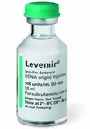
have recently switched one of my canine diabetic patients from NPH insulin to detemir. This is the first time I have used this insulin and I am looking for some help.

Rex is a 10-yr-old, male Schnauzer mix. We started him on 3 units of detemir twice daily and he is currently on 3.5 units twice daily.

We admitted Rex for a glucose curve today. His results were as follows:


8:00 am- 482 mg/dl (fasted and no insulin since 8 pm dose)
8:15 am- 3.5 units of detemir given and dog fed
9:15 am - 445 mg/dl
10:20 am- 301 mg/dl
12:30 pm- 147 mg/dl
1:25 pm- 97 mg/dl
2:30 pm- 80 mg/dl
3:30 pm- 72 mg/dl
4:40 pm- 82 mg/dl
5:10 pm- 108 mg/dl

I like the low glucose values in the middle of the day but am concerned about the severe morning hyperglycemia.

Any recommendations? Thank you for any help.


My Response:

How much does this dog weigh? The dose of detemir (Levemir insulin) we start at is generally around 0.1 mg/kg twice daily, so 7 U per day seems rather high (unless it's a big Schnauzer)

That all said, this curve doesn't look so bad. Like you, I'm not bothered by the afternoon glucoses. They still are normal and not low. Are the owners feeding the same food morning and night? Did the dog eat normally on the day of the glucose curve when hospitalized? If not, that could explain why the insulin worked so well that day; could it be that at home, the glucoses remain higher?

It seems unlikely that the blood sugar would go from 108 mg/dl at 5:10 PM to levels in the 400s 3 hours later when it's time for the shot again. Are the owners giving the insulin and then waiting before feeding?

If the dog is eating well, I like to give the insulin about 30 minutes before eating to ensure some insulin has been absorbed before feeding. We could consider that in this patient if not already being done.

Finally, is this dog feeling well? How if the thirst, appetite and attitude? We must always remember to look at the dog and talk to the owner. We can never just depend on blood glucose curves to make insulin dose adjustments,

Follow-up Information and Additional Questions:

Rex weighs 39 pounds (17.7 kg). The owners report that he is feeling great at home, has gained a little weight recently (1 pound over last 2 months). He is indeed feeling better, playing, and acting normal.

He didn't eat well while here for the curve so I would assume those middle-of-the-day glucose values would be higher at home. I can question the owners better about the timing of the feeding at home. I didn’t realize that could even be a factor!

Do you have owners feed the animal at home before coming in for the curve? Would the next step be to redo the glucose curve on a day when he has eaten that morning?

Would increasing the dose up to 4 units BID before the next curve be advisable?

My Response:

A common problem with the glucose curves is that dogs (or cats) don't eat well in the hospital, and results of a glucose curve really don’t mean much when they haven't eaten. I know it's nice to have a pretreatment glucose, but that's not as important to what else is going on throughout the day.

Given that the dog is doing well at home (which is much more important than ANY blood glucose curve!), I would not raise the dose just yet.

I would recommend repeat the glucose curve following this protocol: I would have them give the insulin injection, wait 15-30 minutes, fed exactly the food and amount that they normally do at home, and then bring the dog in to start the glucose curve shortly thereafter. If the glucose readings drop into the low-normal range as they did before, it would be helpful if you could go longer than 8 hours with your glucose monitoring.


1. Owner gives the insulin injection
2. Wait 30 minutes before feeding
3. Feed exactly the normal diet in the exact amount normally feed every other day
4. Then bring the dog to your hospital to start the glucose curve shortly thereafter
5. Measure blood glucose concentrations every 2 hours for at least 8 hours, but 12 hours is better if possible.

Finally, another way to get around some of these problems is to have the owners do home glucose monitoring. With a bit of training, most dog and cat owners can be trained how to do the measurements at home. This is particularly helpful in cats, where they almost all develop ‘stress’ hyperglycemia in the hospital.

Źródło: endocrinevet.blogspot.com

I have a 14-year-old female spayed DSH feline weighing 5.4 kg, who was diagnosed with diabetes one year ago. To date, she has been receiving 4 IU of PZI (ProZinc) twice daily, and the owners have been very good about keeping her on a diet of high protein/low carbohydrate canned food only.

The owners would like to try switching from the insulin to an oral hypoglycemic agent (glipizide or glyburide). They travel a lot and have difficulty finding someone to look after her, and even having to board her at the clinic is a problem because she is very fractious and difficult to catch and give injections too. The owners themselves can manage just fine with the insulin injections, but no one else can.

Recently, I did some blood work on her and she is doing great. She is no longer polydipsic or polyuric, her serum glucose was normal at 122 mg/dl, and her serum fructosamine was 275 µmol/l (reference range 150-350 µmol/l).

My questions are the following:


- How do I properly transition her to the glipizide or glyburide if we decide to go that route?
- Does this cat even need to continue with insulin or oral glucose products, or can I control her on diet therapy alone at this point?
- Could this cat be going into diabetes remission?


My Response:

The lack of clinical signs and normal fructosamine certainly indicates good diabetes control, and it is possible that the cat is going into remission. Clinical remission is actually not infreqent in cats with well-controlled diabetes mellitus (1).

However, remission, when it does occur, generally develops earlier than a year (generally within the first 3 months of starting insulin therapy) so it's less likely that a cat treated with insulin for a year would be in remission. In addition, when most cats are going into remission, they're generally not normal on 4 units BID — that would be an overdose in most cats in remission. The more typical scenario in a cat going into remission is that their glucose values are dropping too low even on 1 unit BID.

That all said, diabetic remission is possible at any time and it's still possible that your cat is going into remission.

Ideally, the first step in judging remission would be for you would do a complete glucose curve to see if the blood glucose concentrations remain normal through out the day. The finding of a single "normal" blood sugar really doesn't tell you very much at all.

I understand that hospitalizing this fractious cat for a glucose curve would probably not be a pleasant experience for either you or the cat; more importantly, the "stress" would likely make the results meaningless anyway. Can the owners do home glucose monitoring? If so, that would be extremely helpful. Can they, at the very least, measure urine glucose at home?

In the end, how you decide whether the diabetes is going into remission or not is to slowly taper down the insulin dosage by 0.5 U decrements (e.g., start by decreasing from 4 U, BID to 3.5 U, BID). If the blood and/or urine glucose concentration remain normal after a week, then the insulin taper can continue until the insulin can hopefully be discontinued.

The chance of an oral hypoglycemic agent (glibizide or glyburide) working is very small. I personally think it's easier to give insulin to a fractious cat than to give a pill, especially in the hospital or in a kennel.

Remember, oral hypoglycemic agents act by stimulating insulin secretion from the pancreatic islet cells (2). In a cat with borderline diabetes that is going into remission, changing from insulin therapy to one of these drugs may actually be the worst thing to do.

Use of these oral hypoglycemic drugs, by overstimulating any remaining insulin secretion, my actually lead to the final "burn out" of insulin secretion and result in permanent diabetes (2).

Reference:

1. Zini E, Hafner M, Osto M, et al. Predictors of clinical remission in cats with diabetes mellitus. Journal of Veterinary Internal Medicine 2010;24:1314-132
2. Hoenig M, Hall G, Ferguson D, et al. A feline model of experimentally induced islet amyloidosis. American Journal of Pathology 2000; 157:2143-50

Źródło: endocrinevet.blogspot.com

I know that many of you are very busy and may have trouble keeping up with the latest research studies and publications on issues concerning companion animal endocrinology. Therefore, I’ve compiled a fairly extensive list of some of the best clinical endocrine papers written last year (in 2010), and I’ll be sharing these with you over the next few weeks.

We are going to start off with papers that deal with the theme of Diabetes mellitus and its diagnosis and treatment in dogs and cats.

Listed below are 29 research papers written in 2010 that deal with a variety of diabetic topics and issues.

These range from the use of continuous glucose monitoring systems (1, 15, 22) to home glucose monitoring (5, 28); from insulin autoantibodies (4, 18) to insulin resistance (11, 25, 26); from new insulin analogues (8, 10, 24) to diabetic cataracts (12, 21); and from the causes of secondary diabetes (2, 6, 13, 14, 16) to diabetic remission (29).

2010 Papers on Canine and Feline Diabetes Mellitus:

1. Affenzeller N, Benesch T, Thalhammer JG, et al. A pilot study to evaluate a novel subcutaneous continuous glucose monitoring system in healthy Beagle dogs. Veterinary Journal 2010;184:105-110.
2. 3Blois SL, Dickie EL, Kruth SA, et al. Multiple endocrine diseases in cats: 15 cases (1997-2008). Journal of Feline Medicine and Surgery 2010;12:637-642.
3. Claus MA, Silverstein DC, Shofer FS, et al. Comparison of regular insulin infusion doses in critically ill diabetic cats: 29 cases (1999-2007). Journal of Veterinary Emergency and Critical Care 2010;20:509-517.
4. Davison LJ, Herrtage ME, Catchpole B. Autoantibodies to recombinant canine proinsulin in canine diabetic patients. Research in Veterinary Science 2010.
5. Dobromylskyj MJ, Sparkes AH. Assessing portable blood glucose meters for clinical use in cats in the United Kingdom. The Veterinary Record 2010;167:438-442.
6. Fall T, Hedhammar A, Wallberg A, et al. Diabetes mellitus in elkhounds is associated with diestrus and pregnancy. Journal of Veterinary Internal Medicine 2010;24:1322-1328.
7. Furrer D, Kaufmann K, Reusch CE, et al. Amylin reduces plasma glucagon concentration in cats. Veterinary Journal 2010;184:236-240.
8. Gilor C, Graves TK. Synthetic insulin analogs and their use in dogs and cats. The Veterinary Clinics of North America Small Animal Practice 2010;40:297-307.
9. Gilor C, Graves TK, Lascelles BD, et al. The effects of body weight, body condition score, sex, and age on serum fructosamine concentrations in clinically healthy cats. Veterinary Clinical Pathology 2010;39:322-328.
10. Gilor C, Ridge TK, Attermeier KJ, et al. Pharmacodynamics of insulin detemir and insulin glargine assessed by an isoglycemic clamp method in healthy cats. Journal of Veterinary Internal Medicine 2010;24:870-874.
11. Hess RS. Insulin resistance in dogs. The Veterinary Clinics of North America Small Animal Practice 2010;40:309-316.
12. Kador PF, Webb TR, Bras D, et al. Topical KINOSTAT ameliorates the clinical development and progression of cataracts in dogs with diabetes mellitus. Veterinary Ophthalmology 2010;13:363-368.
13. McLauchlan G, Knottenbelt C, Augusto M, et al. Retrospective evaluation of the effect of trilostane on insulin requirement and fructosamine concentration in eight diabetic dogs with hyperadrenocorticism. The Journal of Small Animal Practice 2010;51:642-648.
14. Meij BP, Auriemma E, Grinwis G, et al. Successful treatment of acromegaly in a diabetic cat with transsphenoidal hypophysectomy. Journal of Feline Medicine and Surgery 2010;12:406-410.
15. Moretti S, Tschuor F, Osto M, et al. Evaluation of a novel real-time continuous glucose-monitoring system for use in cats. Journal of Veterinary Internal Medicine 2010;24:120-126.
16. Niessen SJ. Feline acromegaly: an essential differential diagnosis for the difficult diabetic. Journal of feline medicine and surgery 2010;12:15-23.
17. Niessen SJ, Powney S, Guitian J, et al. Evaluation of a quality-of-life tool for cats with diabetes mellitus. Journal of Veterinary Internal Medicine 2010;24:1098-1105.
18. Nishii N, Takasu M, Kojima M, et al. Presence of anti-insulin natural autoantibodies in healthy cats and its interference with immunoassay for serum insulin concentrations. Domestic Animal Endocrinology 2010;38:138-145.
19. Nishii N, Yamasaki M, Takasu M, et al. Plasma leptin concentration in dogs with diabetes mellitus. The Journal of Veterinary Medical Science 2010;72:809-811.
20. O'Brien MA. Diabetic emergencies in small animals. The Veterinary Clinics of North America Small Animal Practice 2010;40:317-333.
21. Oliver JA, Clark L, Corletto F, et al. A comparison of anesthetic complications between diabetic and nondiabetic dogs undergoing phacoemulsification cataract surgery: a retrospective study. Veterinary Ophthalmology 2010;13:244-250.
22. Reineke EL, Fletcher DJ, King LG, et al. Accuracy of a continuous glucose monitoring system in dogs and cats with diabetic ketoacidosis. Journal of Veterinary Emergency and Critical Care 2010;20:303-312.
23. Rucinsky R, Cook A, Haley S, et al. AAHA diabetes management guidelines. Journal of the American Animal Hospital Association 2010;46:215-224.
24. Sako T, Mori A, Lee P, et al. Time-action profiles of insulin detemir in normal and diabetic dogs
25. Scott-Moncrieff JC. Insulin resistance in cats. The Veterinary Clinics of North America Small Animal Practice 2010;40:241-257.
26. Verkest KR, Fleeman LM, Rand JS, et al. Basal measures of insulin sensitivity and insulin secretion and simplified glucose tolerance tests in dogs. Domestic Animal Endocrinology 2010;39:194-204.
27. Zeugswetter F, Handl S, Iben C, et al. Efficacy of plasma beta-hydroxybutyrate concentration as a marker for diabetes mellitus in acutely sick cats. Journal of Feline Medicine and Surgery 2010;12:300-305.
28. Zeugswetter FK, Rebuzzi L, Karlovits S. Alternative sampling site for blood glucose testing in cats: giving the ears a rest. Journal of Feline Medicine and Surgery 2010;12:710-713.
29. Zini E, Hafner M, Osto M, et al. Predictors of clinical remission in cats with diabetes mellitus. Journal of Veterinary Internal Medicine 2010;24:1314-1321.

Źródło: endocrinevet.blogspot.com

My patient is a 4-year-F/S Shetland sheepdog who has been treated with melatonin and flax seed oil since she was 11-months-old. Her main clinical sign was a chronic decrease in appetite, which started at 8-months of age when she had her first heat cycle.

Vaginal smears and cytology at that time was consistent with early estrus. She did not have noticeable bleeding, but she is very "tidy" and licks herself a lot. No polydipsia or polyuria was noted, and her hair coat has always been completely normal. Results of her physical examination were completely normal.

Two months later, we decided to spay her in case there was any connection between going into heat and the lack of appetite. A CBC and serum chemistry panel done before surgery were both normal. Routine ovariohysterectomy was performed. Examination of the uterus showed that it was more turgid than normal, but it did not show any evidence for pyometra or mucometra.

After surgery the dog became totally anorexic and vomited multiple time. She was referred to a local internal medicine specialist for workup. Based upon results of abdominal ultrasound, endoscopic exam and intestinal/stomach biopsies, a diagnosis of helicobacter pylori infection and inflammatory bowl disease (IBD) was made. Treatment with amoxicillin, metronidazole, and a food allergy diet was instituted, which quickly cleared up the intestinal signs and anorexia.

Because of the finding of bilateral adrenomegaly on abdominal ultrasound, however, an ACTH stimulation test was also performed shortly after surgery. Serum samples for an adrenal panel were submitted to the University of Tennessee Clinical Endocrinology Service Laboratory.

Atypical Cushing's disease was diagnosed on the basis the finding of a high baseline estradiol concentration (105.1 pg/ml; reference range, 30-69. pg/ml) as well as a high ACTH-stimulated estradiol value (111.1 pg/ml; 28-69 pg/ml). The other sex steroids tested (including 17-hydroyxprogrogesteone) were all within normal limits. Based on these results, she was started melatonin and flax seed oil for the atypical Cushing's syndrome.

Now 3 years later, the owner is asking me if her dog really needs the melatonin and flax seed oil. She has declined the offer of retesting the sex steroid panel on several occasions. The owner says her dog has been normal in every way, except for mild lethargy during the morning after she gives the melatonin. The owner believes the melatonin is causing sleepiness.

My questions are:

1. Do you think this dog has atypical Cushing's disease? It doesn't appear that she ever had any clinical signs of the disease.
2. If we stop the melatonin, how would we determine the need to go back on the drug?

My Response:

I really must admit that I've heard it all now — diagnosing atypical Cushing's disease in an 11-month old dog with the primary complaints of anorexia, inflammatory bowl disease, and helicobacter!

You cannot base a diagnosis of Cushing's disease — typical or atypical — on the finding of large adrenal glands on an ultrasound exam. This dog was severely ill when the ultrasound and ACTH stimulation test were performed. One would expect a sick dog to have a "stress" response, which would include increased secretion of pituitary ACTH leading to increased cortisol section. With chronic illness and continued stress, bilateral adrenocortical hyperplasia would be an expected finding as cortisol hypersecretion continues.

Cushing's syndrome (either typical or atypical disease) is a clinical diagnosis and must be based primarily on the dog's clinical features and physical examination findings. Dogs with Cushing's syndrome do NOT have anorexia and vomiting. They have a good to increased appetite, together with polyuria, polydipsia, hair loss, potbelly, and weakness. In my opinion, it was totally inappropriate to even test the dog for atypical Cushing's disease, given the total lack of any clinical features of the disease. Plus, this disease develops in older dogs, not dogs that are less than a year of age.

Bottom line:


- In any dog with chronic nonadrenal disease, the finding of enlarged adrenal glands with abdominal ultrasonography is not an uncommon finding due to the chronic stress of illness.
- This finding of "big adrenals" can never be used to confirm a diagnosis of Cushing's syndrome — be it typical or atypical hyperadrenocorticism.
- Diagnosis must be based on a combination of the typical clinical features of Cushing's disease together with results of standard pituitary function testing (i.e., low-dose dexamethasone suppression and ACTH stimulation testing).

I agree totally with the owner here. I would stop the melatonin and flax seed oil and monitor the clinical response. Why monitor for hormones when we don't even know what the elevations mean? We see many dogs tested with the sex steroid panel that have high serum estradiol values when the other hormone concentrations are normal.

In fact, I rarely see ANY dog tested that has normal values for estradiol. I can only assume that the labor seen a high incidence of false-positive results.

In support of my clinical experience, a recent research paper published by the head dermatologist at the University of Tennessee, College of Veterinary Medicine showed that serum estradiol concentrations varied considerably in clinically normal dogs (1). They concluded in this study that the finding of high estradiol concentrations alone cannot be used to diagnose atypical Cushing's syndrome, because of the high variability of these hormone results.

References:

Frank LA, Mullins R, Rohrbach BW. Variability of estradiol concentration in normal dogs. Vet Dermatology. 2010; 21:490-493.

Źródło: endocrinevet.blogspot.com

3

bony.jpg

zoetis2024

megavet